Figures & data
Figure 1 A schematic illustration to show the formation of I-TMSG complex, as well as its theranostic application to photothermal therapy treatment and near-infrared fluorescence imaging.
Abbreviations: I-TMSG, tLyp-1 peptide-functionalized, indocyanine green-containing mesoporous silica-coated gold nanorods; PEG3500, polyethylene glycol-3500; NIR, near-infrared.
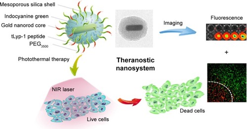
Figure 2 A schematic procedure for the preparation of TMSG nanoparticles.
Abbreviations: TMSG, tLyp-1 peptide-functionalized and polyethylene glycol-modified mesoporous silica-coated gold nanorods; APTES, aminopropyl triethoxysilane; NHS-PEG3500-MAL maleimide PEG N-hydroxylsuccinimide ester; CTAB, cetyltrimethylammonium bromide; h, hours; TEOS, tetraethyl orthosilicate.
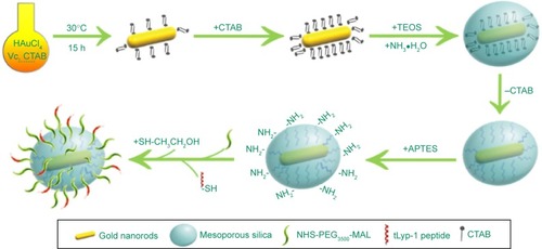
Figure 3 Transmission electron micrograph images of (A) GNRs (inset: the photo of CTAB-stabilized GNRs solution) and (B) MSG. (C) High-magnification transmission electron micrograph image of MSG. (D) UV–vis spectra of GNRs and MSG. (E) N2 adsorption–desorption isotherms. (F) The pore diameter distribution of MSG.
Abbreviations: GNRs, gold nanorods; CTAB, cetyltrimethylammonium bromide; MSG, mesoporous silica-coated GNRs; UV, ultraviolet; SPT, standard pressure and temperature.
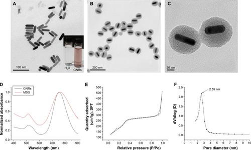
Figure 4 (A) Dynamic light scattering size of MSG. (B) Zeta potential of GNRs (CTAB stabilized), MSG, MSG-NH2, PMSG, and TMSG (n=3). (C) X-ray photoelectron spectra of MSG and TMSG. (D) TGA curves of MSG, PMSG, and TMSG.
Abbreviations: MSG, mesoporous silica-coated GNRs; GNRs, gold nanorods; CTAB, cetyltrimethylammonium bromide; MSG-NH2, amine-functionalized MSG; PMSG, polyethylene glycol-modified MSG; TMSG, tLyp-1 peptide-functionalized PMSG; s, second; TGA, thermogravimetric analysis.
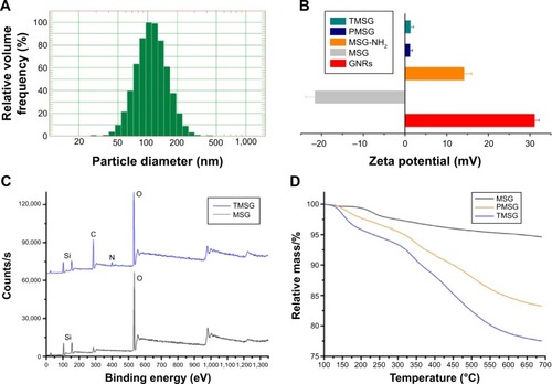
Figure 5 The UV–vis (A–C) or FL (D) spectra of I-TMSG and ICG solutions at different points.
Note: The results demonstrated that the probe had a high stability compared to freely dissolved ICG and the fluorescence quenching of ICG encapsulated intensively in the mesoporous of TMSG.
Abbreviations: UV, ultraviolet; FL, fluorescence; I-TMSG, tLyp-1 peptide-functionalized, indocyanine green-containing mesoporous silica-coated gold nanorods; ICG, indocyanine green.

Figure 6 (A) The FL image and (B) relative FL intensity of I-TMSG nanoparticles or free ICG in culture medium coincubated with MDA-MB-231 cells or without cells at different points in time, respectively.
Abbreviations: FL, fluorescence; I-TMSG, tLyp-1 peptide-functionalized, indocyanine green-containing mesoporous silica-coated gold nanorods; ICG, indocyanine green; MDA-MB-231 cells, MD Anderson-metastatic breast-231 cells; h, hours.
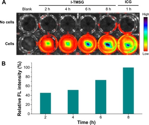
Figure 7 Confocal laser scanning microscope images of MDA-MB-231 cells incubated with rhodamine-labeled PMSG or TMSG. Blue: Hoechst 33342 and red: rhodamine B.
Abbreviations: PMSG, polyethylene glycol-modified mesoporous silica-coated gold nanorods; TMSG, tLyp-1 peptide-functionalized PMSG; h, hours; MDA-MB-231cells, MD Anderson-metastatic breast-231 cells.
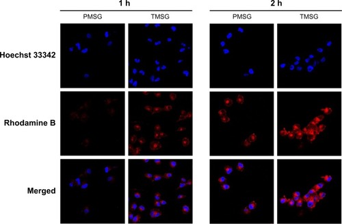
Figure 8 (A) Cellular uptake of rhodamine-labeled PMSG or TMSG in MDA-MB-231 cells and then quantization of mean FL intensity by flow cytometry. (B) Cellular association of rhodamine-labeled TMSG in the presence of different endocytosis inhibitors in MDA-MB-231 cells.
Notes: The geometric mean of the fluorescence for the cells treated with rhodamine-labeled TMSG without any inhibitors was defined as 100% (n=3). ***P<0.001, **P<0.01, *P<0.05, and NS, not statistically significant.
Abbreviations: FL, fluorescence; PMSG, polyethylene glycol-modified mesoporous silica-coated gold nanorods; TMSG, tLyp-1 peptide-functionalized PMSG; min, minutes; M-β-CD, methyl-β-cyclodextrin; MDA-MB-231cells, MD Anderson-metastatic breast-231 cells.
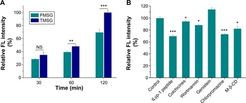
Figure 9 Relative viabilities of MDA-MB-231 cells after being incubated with (A) PMSG and TMSG nanoparticles or (B) I-PMSG and I-TMSG nanoparticles for 48 hours (n=4).
Abbreviations: PMSG, polyethylene glycol-modified mesoporous silica-coated gold nanorods; TMSG, tLyp-1 peptide-functionalized PMSG; I-PMSG, indocyanine green-containing PMSG; I-TMSG, indocyanine green-containing TMSG; MDA-MB-231cells, MD Anderson-metastatic breast-231 cells.
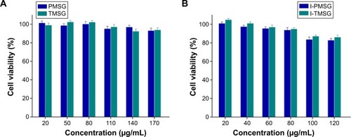
Figure 10 (A) Temperature curves and (B) thermograph map of different concentrations of TMSG under exposure to a 785-nm laser (3 W/cm2) over a period of 2 minutes.
Notes: (a) 60 μg/mL, (b) 40 μg/mL, (c) 20 μg/mL, (d) 10 μg/mL, and (e) 0 μg/mL.
Abbreviations: TMSG, tLyp-1 peptide and polyethylene glycol co-modified mesoporous silica-coated gold nanorods; s, seconds.
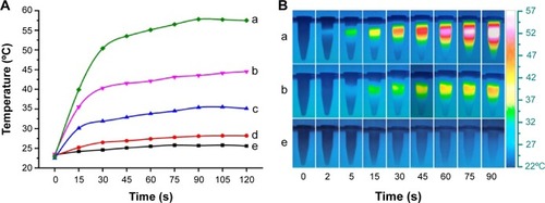
Figure 11 Confocal laser scanning microscope images of Calcein-AM/propidium iodide-costained MDA-MB-231 cells in different treatment conditions.
Notes: The laser irradiated regions are inside a circular area, and the edges are marked with white dashed lines. Viable cells are stained green with Calcein-AM, and dead/later apoptosis cells are floating and eluted or are stained red with propidium iodide.
Abbreviations: DMEM, Dulbecco’s Modified Eagle’s Medium; TMSG, tLyp-1 peptide and polyethylene glycol comodified mesoporous silica-coated gold nanorods; MDA-MB-231cells, MD Anderson-metastatic breast-231 cells.
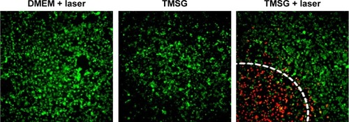
Figure 12 Flow cytometry analysis of MDA-MB-231 cells after photothermal therapy treatments under different treatment conditions.
Notes: The living cell fraction is negative for both Annexin V-FITC and propidium iodide. An earlier stage of apoptosis is linked with positive Annexin V-FITC labeling only. Double-stained cells were considered as necrotic/late apoptotic cells. The concentration of TMSG was 70 μg/mL, and the power density of laser irradiation was 3 W/cm2.
Abbreviations: TMSG, tLyp-1 peptide and polyethylene glycol-comodified mesoporous silica-coated gold nanorods; PI, propidium iodide; FL, fluorescence; MDA-MB-231 cells, MD Anderson-metastatic breast-231 cells; min, minutes.
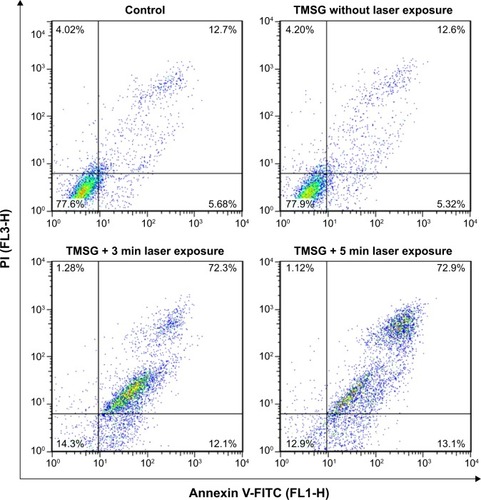
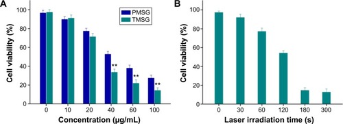
Figure 13 (A) Relative viabilities of MDA-MB-231 cells incubated with TMSG or PMSG at different concentrations under exposure to a 785 nm laser at a power density of 3 W/cm2 for 5 minutes. (B) Relative viabilities of MDA-MB-231 cells after TMSG (80 μg/mL) induced photothermal ablation over different laser exposure periods.
Notes: The cell viability values were all normalized to control untreated cells (n=3). **P<0.01.
Abbreviations: PMSG, polyethylene glycol-modified mesoporous silica-coated gold nanorods; TMSG, tLyp-1 peptide-functionalized PMSG; s, seconds; MDA-MB-231cells, MD Anderson-metastatic breast-231 cells.