Figures & data
Table 1 Summary of the pharmacokinetic parameters of the cholinesterase inhibitors and memantine
Table 2 Summary of nanotechnology-based systems applied in the treatment of Alzheimer’s disease
Figure 1 Formation of amyloid plaques (A) and neurofibrillary tangles (B) in the neurons in Alzheimer’s disease.
Abbreviations: Aβ, β-amyloid; APP, amyloid precursor protein.
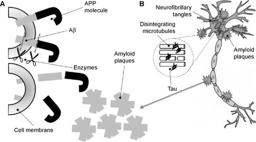
Figure 2 Schematic differences between nanocapsule, nanostructured lipid carrier, polymeric nanoparticle, and solid lipid nanoparticle drug delivery systems.
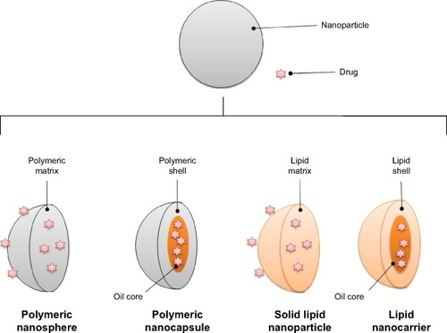
Figure 3 Schematic representation of types of liposomes and enlarged view of the layers of phospholipids.
Abbreviations: GUV, giant unilamellar vesicle; LUV, large unilamellar vesicle; MLV, multilamellar vesicle; SUV, small unilamellar vesicle.
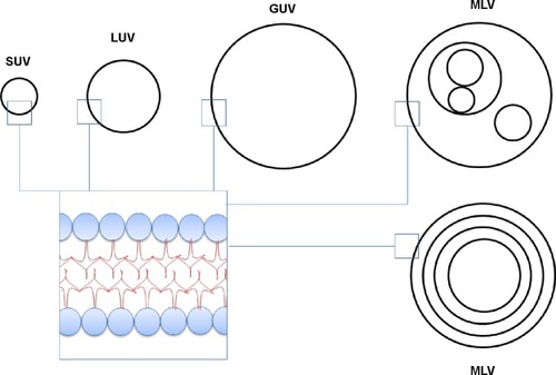
Figure 4 Main pathways for nanosystems to cross the blood–brain barrier to target to brain.
Abbreviations: CNS, central nervous system; NCLs, nanostructured lipid carriers; NPs, nanoparticles.
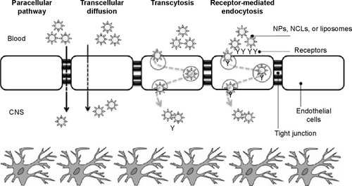
Figure 5 Photograph of microemulsion and nanoemulsion.
Note: Enlarged areas show schematics of the size of droplets formed.
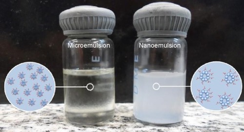
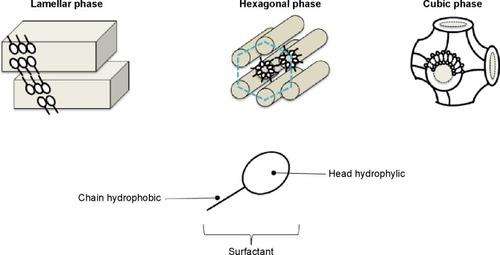
Figure 6 Schematic representation of lamellar, hexagonal, and cubic liquid crystal mesophases formed by surfactant molecules’ self-assembly.