Figures & data
Figure 1 Schematic illustration of the PEGylated QDs-based immunofiltration assay.
Notes: (A) The structure illustration of the immunofiltration pad. (B) The detection process includes an initial step of serum sample mixing with QD-antibody conjugate followed by sample loading and washing with buffer. (C) The qualitative detection is monitored by naked eye under UV light illumination, while the quantitative detection of the resulted fluorescent signals is carried out using an optical fiber spectroscopy and 405 nm laser excitation.
Abbreviations: QD, quantum dot; UV, ultraviolet; NC, nitrocellulose; PEG, polyethylene glycol.
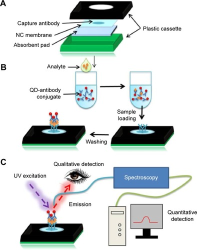
Figure 2 Synthesis and characterization of PEGylated QDs and QD-antibody conjugates.
Notes: (A) Schematic of QD and QD-antibody structure applied for CRP detection using IFA system. Identical fluorescence emission (B) and UV-Vis absorbance (C) spectra of GSH-QDs (black circle) and PEGylated GSH-QDs (red rhombus) dissolved in PBS buffer solution (0.01 M, pH 7.2). (D) Agarose gel electrophoresis of QDs and QD-antibody conjugates, running at 80 V for 20 minutes.
Abbreviations: Ab, antibody; QD, quantum dot; UV-Vis, ultraviolet-visible; IFA, immunofiltration assay; CRP, C-reactive protein; GSH, glutathione; PEG, polyethylene glycol; PL, photoluminescence; PBS, phosphate-buffered saline.
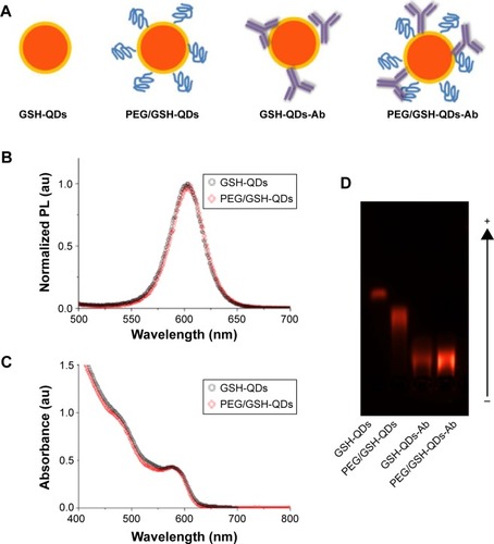
Figure 3 Comparative detection of CRP using QDs labeling conjugates prepared from GSH-QDs and PEG/GSH-QDs conjugates.
Note: 5 µL of standard CRP sample in serum added to 200 µL of 20 nM QD conjugates solution, then 120 µL aliquot was loaded into IFA pad.
Abbreviations: QD, quantum dot; IFA, immunofiltration assay; CRP, C-reactive protein; GSH, glutathione; PEG, polyethylene glycol.
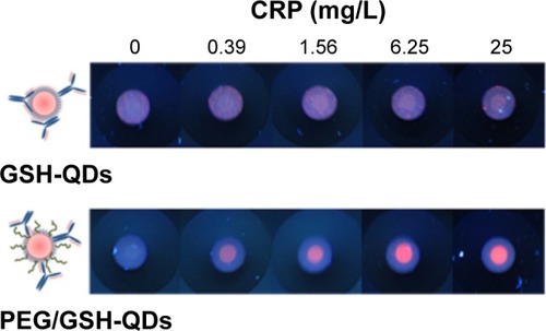
Figure 4 Optimization of the detection conditions for PEGylated QDs-based immunofiltration assay for CRP.
Notes: Typical images (A) and quantitative fluorescent response (B) of QDs-base immunofiltration assay for negative (0 mg/L) and positive (0.5 mg/L) CRP samples in serum using different concentrations of QDs conjugates. The inset figure in (B) shows the signal/noise (S/N) ratio calculated from the PL intensity ratio of positive to negative samples under different concentrations of the QDs conjugates. (5 µL of CRP samples in serum added to 200 µL of different concentration of QD conjugates solution, then 120 µL aliquot was loaded into IFA pad.)
Abbreviations: QD, quantum dot; Ab, antibody; IFA, immunofiltration assay; CRP, C-reactive protein; PEG, polyethylene glycol; PL, photoluminescence.
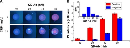
Figure 5 Fluorescent images (upper) and spectra (lower) of immunofiltration pad spot tested with a series of CRP samples.
Notes: 5 µL of CRP samples in serum respectively added to 400 µL (A) and 800 µL (B) of 20 nM QD conjugates solution, then 120 µL aliquot was loaded into IFA pad.
Abbreviations: QD, quantum dot; IFA, immunofiltration assay; CRP, C-reactive protein; PL, photoluminescence.

Figure 6 The quantitative dynamic range of the developed QDs-based immunofiltration assay using different volume of QDs conjugates.
Abbreviations: QD, quantum dot; CRP, C-reactive protein; PL, photoluminescence.
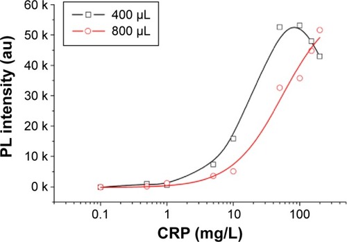
Figure 7 Application of the developed QD-based immunofiltration assay for CRP detection.
Notes: (A) Typical fluorescent images of detection results carried out with twofold serially diluted CRP calibrator and a negative control (0 mg/L CRP serum sample) 5 µL of CRP samples in serum respectively added to 800 µL of 20 nM QD conjugates solution, then 120 µL aliquot was loaded into IFA pad. (B) The calibration curve of the quantitative detection by the developed QD-based immunofiltration assay. (C) Correlation between the results of QD-based immunofiltration assay and latex enhanced immune-agglutination assay for 50 human serum samples.
Abbreviations: QD, quantum dot; IFA, immunofiltration assay; CRP, C-reactive protein; PL, photoluminescence.
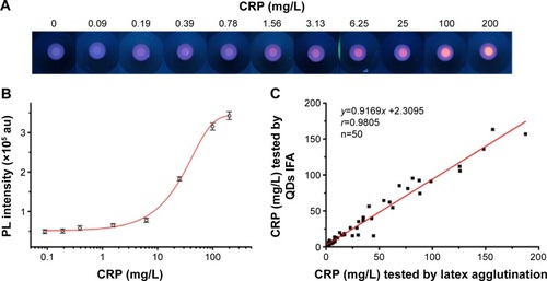
Table 1 Comparison of developed QD-based IFA with other QDs-based assays and traditional ELISA methods for CRP detection
Figure S1 UV-Vis absorbance and fluorescent spectra of CdSe core and CdSe/ZnSe/ZnS core/shell/shell QDs.
Abbreviations: UV, ultraviolet; UV-Vis, ultraviolet-visible; PL, photoluminescence; QDs, quantum dots.
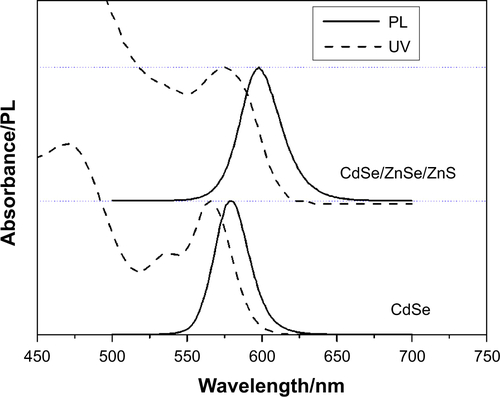
Figure S2 TEM graphs of CdSe core (A) and CdSe/ZnSe/ZnS core/shell/shell (B) QDs.
Abbreviations: TEM, transmission electron microscopy; QDs, quantum dots.
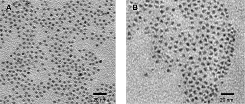
Figure S3 Dynamic light scattering analysis of GSH-QDs.
Abbreviations: QDs, quantum dots; GSH, glutathione.
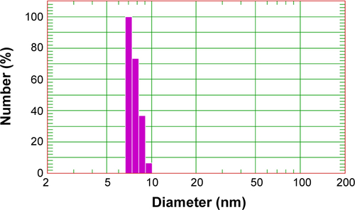
Figure S4 Dynamic light scattering analysis of PEG/GSH-QDs.
Abbreviations: QDs, quantum dots; GSH, glutathione; PEG, polyethylene glycol.
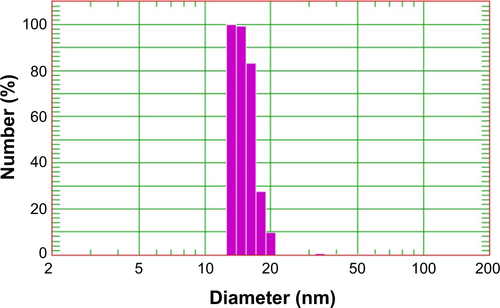
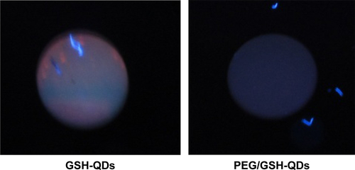
Figure S5 Comparison of non-specific binding of GSH-QDs and PEG/GSH-QDs conjugates with NC membrane without capturing antibody.
Abbreviations: QD, quantum dot; GSH, glutathione; PEG, polyethylene glycol; NC, nitrocellulose.