Figures & data
Figure 1 The MRI signal alterations in liver and spleen.
Notes: (A and B) are the MRI images before and after ATF-IONP injection, respectively, without and with PEG coating, both of them are phased from up to down. (C and D) are the variations of T2 signal intensity; (E and F) are the variations of T1 signal intensity. Monkey 1 received ATF-IONP, and Monkey 2 received ATF-PEG-IONP.
Abbreviations: ATF, amino-terminal fragment; IONP, magnetic iron oxide nanoparticle; MRI, magnetic resonance imaging; PEG, polyethylene glycol.
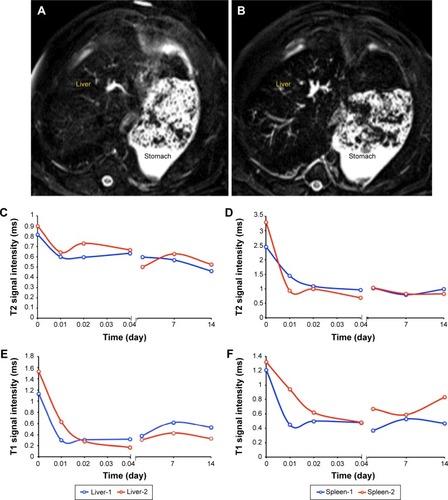
Figure 2 Plots of time-dependent changes of IONP contents in rhesus monkeys (A–I).
Notes: The plots suggest the effects of ATF-IONPs as indicated by serological examination. The gray areas show the normal reference range provided by Sichuan Primed Bio-Tech Group Co, Ltd, Chengdu, People’s Republic of China.
Abbreviations: ALP, alkaline phosphatase; ALT, alanine transaminase; AST, aspartate transaminase; BIL-D, direct bilirubin; CK, creatine kinase; CREA, creatinine; HGB, hemoglobin; RBC, red blood cell; WBC, white blood cell; ATF, amino-terminal fragment; IONPs, magnetic iron oxide nanoparticles.
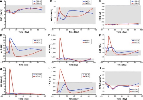
Figure 3 Determination of PK of uPAR-targeted IONPs in rhesus monkeys.
Notes: After the administration of IONPs, the concentrations of probes change with time elapsed. Upper panel: A monkey received 5 mg/kg of ATF-IONP. Lower panel: A monkey received 5 mg/kg of ATF-PEG-IONP.
Abbreviations: ATF, amino-terminal fragment; AUC, area under the curve; Cl_obs, systemic clearance; C0, initial concentration extrapolated to time zero; Cmax, maximum serum concentration; IONPs, magnetic iron oxide nanoparticles; PEG, polyethylene glycol; PK, pharmacokinetics; MRT, mean residence time; T1/2, half-life; uPAR, urokinase plasminogen activator receptor; Vss_obs, apparent volume of distribution at steady state.
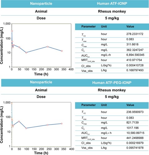
Figure 4 Detection of IONPs in the livers of rhesus monkeys by Prussian Blue staining.
Notes: Original magnification: ×200; magnified to ×400 in black box. (A and C) are the biopsy samples of the liver obtained at 24 hours and massive IONPS seen in the kupffer cells (+ + +), and (B and D) are samples obtained at 3 months after the IONP injection, and sparse IONPs seen in the kupffer cells (+). The liver of Monkey 2 has weaker blue staining compared with that of Monkey 1 (A and C, respectively). And the levels of IONPs in the liver of monkeys are significantly decreased at 3 months posttreatment. No significant difference between (B and D) was observed.
Abbreviation: IONPs, magnetic iron oxide nanoparticles.
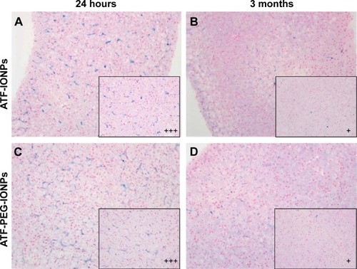
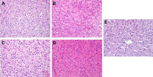
Figure 5 Effects of IONPs in the liver of the rhesus monkeys by H&E staining.
Notes: Original magnification: ×400. (A and B) are the liver samples of Monkey 1 at 24 hours and 3 months posttreatment, respectively. (C and D) are the liver samples of Monkey 2 at 24 hours and 3 months posttreatment, respectively. Obvious swelling of hepatocytes and minimal necrosis were seen at 24-hour posttreatment stage, as well as recovery to normal with little swelling of hepatocytes in Monkey 1 at the 3-month posttreatment stage, compared with the negative control. No significant differences between (A and C) and between (B and D) were observed. (E) is the negative control.
Abbreviations: IONPs, magnetic iron oxide nanoparticles; H&E, hematoxylin–eosin.