Figures & data
Figure 1 Characterization of three rod-like gold-mesoporous silica nanoparticles.
Notes: TEM images showing bare AuNPs (A), core–shell Au@mSiO2NPs (B), and Janus Au@mSiO2NPs (C). The scale bar represents 200 nm.
Abbreviations: AuNPs, gold nanoparticles; TEM, transmission electron microscopy.
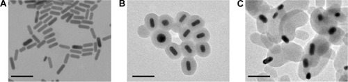
Figure 2 Endocytosis of three rod-like gold-mesoporous silica nanoparticles.
Notes: Confocal microscopy of the cellular uptake behavior of FITC-labeled AuNPs, core–shell Au@mSiO2NPs, and Janus Au@mSiO2NPs in MCF-7 cells. Green florescence, FITC; blue florescence, nuclei stained with Hoechst 33258. Scale bars represent 10 µm.
Abbreviations: AuNPs, gold nanoparticles; FITC, fluorescein isothiocyanate.
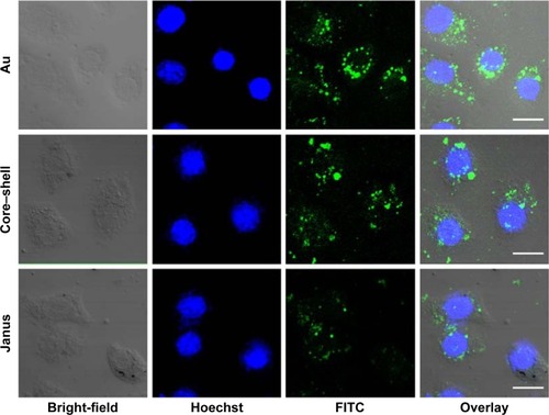
Figure 3 Mitochondrial apoptosis induced by three rod-like gold-mesoporous silica nanoparticles.
Notes: (A) MTT assay showing MCF-7 cells after incubation with three rod-like gold-mesoporous silica nanoparticles at various concentrations (1.56–200 µg/mL) for 24 hours. (B) MTT assay showing MCF-7 cells after separate incubation with 12.5 µg/mL of three rod-like gold-mesoporous silica nanoparticles for various time intervals (24, 48, or 72 hours). (C) MCF-7 cells were treated separately with 12.5 µg/mL of three rod-like gold-mesoporous silica nanoparticles for 24 hours; the apoptotic ratio was then assessed by Annexin V-FITC/PI binding and measured using flow cytometry. (D) Changes in the MMP were detected using JC-1 staining and analyzed by flow cytometry. The data represent three separate experiments. Mean values ± SD. *P<0.05 versus a control group, **P<0.01 versus a control group.
Abbreviations: Au, gold; FITC, fluorescein isothiocyanate; PI, propidium iodide; MTT, 3-(4,5-dimethylthiazol-2-yl)-2,5-diphenyltetrazolium bromide.
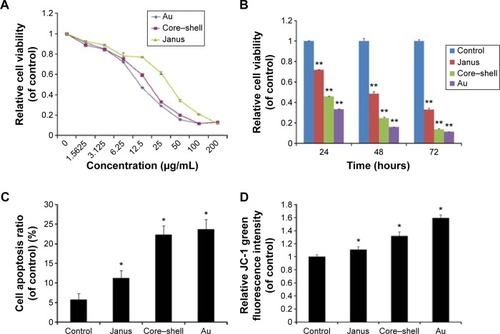
Figure 4 ROS generation induced by three rod-like gold-mesoporous silica nanoparticles.
Notes: MCF-7 cells were separately treated with 12.5 µg/mL of three rod-like gold-mesoporous silica nanoparticles for 24 hours; then, intracellular ROS were stained using DCFH-DA and measured using fluorescence microscopy (A) and flow cytometry (B). The scale bars represent 50 µm. The data represent three separate experiments. Mean values ± SD. *P<0.05 versus a control group.
Abbreviations: AuNPs, gold nanoparticles; ROS, reactive oxygen species; DCFH-DA, 2′7′-dichlorofluorescein diacetate; SD, standard deviation.
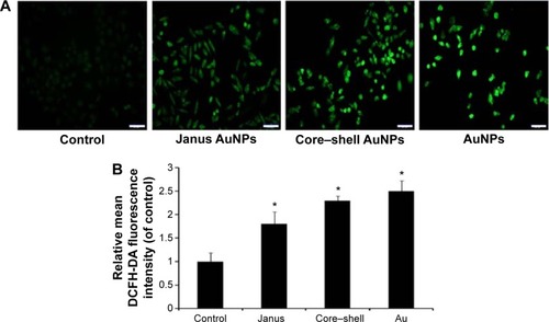
Figure 5 Roles played by MAPK subfamilies in the mitochondrial apoptosis induced by three rod-like gold-mesoporous silica nanoparticles.
Notes: MCF-7 cells were separately treated with 12.5 µg/mL of three rod-like gold-mesoporous silica nanoparticles for 24 hours; then, whole-cell lysates were collected and immunoblotted using antibodies against MAPK subfamily proteins, such as ERK, p-ERK, p38, p-p38, JNK, and p-JNK (A), as well as apoptosis-related proteins, such as cyt c, Bcl-2, Bax, caspase-9, caspase-3, and PARP (B). GAPDH was used as a standard to ensure the equal loading of lysates. (C) Quantitative analysis of the ratio of Bax/Bcl-2. Values are presented as means ± SD of three determinations. *P<0.05 versus control group.
Abbreviations: Au, gold; MAPK, mitogen-activated protein kinase; ERK, extracellular-signal-regulating kinase; JNK, c-Jun-N-terminal kinase; cyt c, cytochrome c; Bcl-2, B-cell lymphoma-2; Bax, Bcl-2-associated X protein; PARP, polyADP-ribose polymerase; GAPDH, glyceraldehyde-3-phosphate dehydrogenase; SD, standard deviation.
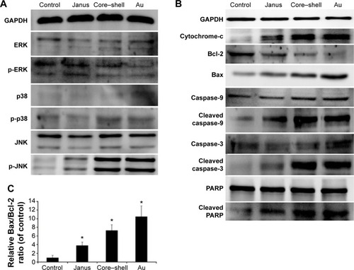
Figure 6 The effect of ROS on the mitochondrial apoptosis induced by three rod-like gold-mesoporous silica nanoparticles.
Notes: MCF-7 cells were separately treated with 12.5 µg/mL of three rod-like gold-mesoporous silica nanoparticles for 24 hours in the presence or absence of 5 mM NAC. (A) Cytotoxicity was analyzed using an MTT assay. (B) The apoptotic ratio was assessed based on Annexin V-FITC/PI binding and measured using flow cytometry analysis. (C) Intracellular ROS were stained using DCFH-DA and measured using flow cytometry. (D) The expression of p-JNK and cleaved caspase-3 was analyzed using western blotting. The data represent three separate experiments. Mean values ± SD. *P<0.05 versus a control group, **P<0.01 versus a control group.
Abbreviations: Au, gold; DCFH-DA, 2′7′-dichlorofluorescein diacetate; ROS, reactive oxygen species; FITC, fluorescein isothiocyanate; PI, propidium iodide; MTT, 3-(4,5-dimethylthiazol-2-yl)-2,5-diphenyltetrazolium bromide; NAC, N-acetylcysteine; JNK, c-Jun-N-terminal kinase; SD, standard deviation.
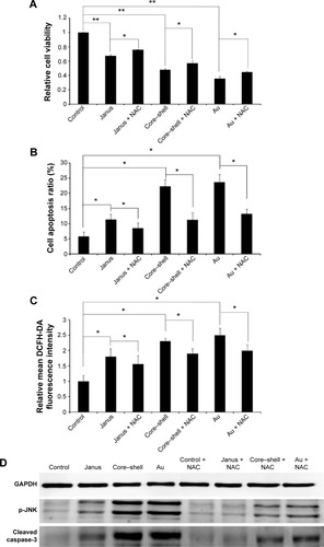
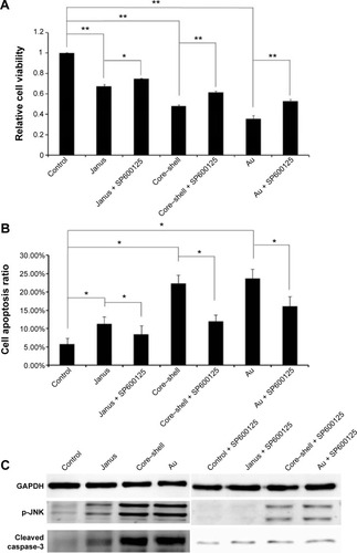
Figure 7 The effect of JNK on the mitochondrial apoptosis induced by three rod-like gold-mesoporous silica nanoparticles.
Notes: MCF-7 cells were separately treated with 12.5 µg/mL of three rod-like gold-mesoporous silica nanoparticles for 24 hours in the presence or absence of 30 µM SP600125. (A) Cytotoxicity was analyzed using an MTT assay. (B) Then, the apoptotic ratio was assessed based on Annexin V-FITC/PI binding and measured using flow cytometry. (C) The expression of p-JNK and cleaved caspase-3 was analyzed using western blotting. The data represent three separate experiments. Mean values ± SD. *P<0.05 versus a control group, **P<0.01 versus a control group.
Abbreviations: Au, gold; FITC, fluorescein isothiocyanate; GAPDH, glyceraldehyde-3-phosphate dehydrogenase; PI, propidium iodide; MTT, 3-(4,5-dimethylthiazol-2-yl)-2,5-diphenyltetrazolium bromide; SP600125, 1,9-pyrazoloanthrone; JNK, c-Jun-N-terminal kinase; SD, standard deviation.