
Figures & data
Figure 1 Schematic illustration of the geometry of multifunctional GQDs for the traceable, targeted delivery of anticancer drugs and their interactions with cancer cells.
Abbreviations: Dox, doxorubicin; EDC/NHS, 1-(3-(dimethylamino)propyl)-3-ethylcarbodiimide and N-hydroxysuccinimide; GQD, graphene quantum dot; RGD, arginine-glycine-aspartic acid.
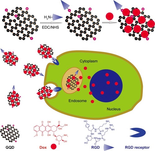
Figure 2 Physicochemical characterization of GQDs.
Notes: (A) GQDs dispersed in PBS and illuminated with sunlight (left) and UV light (right). (B) PL spectrum of GQDs excited at wavelengths from 365 to 543 nm. (C) PL spectrum of GQDs under different pH values. (D) Transmission electron microscope image of GQDs. Insets are high-resolution transmission electron microscopy image (left) and the corresponding fast Fourier transform pattern (right). (E) Lateral size distribution of GQDs. (F) Fourier transform infrared spectrum of GQDs.
Abbreviations: GQDs, graphene quantum dots; PBS, phosphate-buffered saline; PL, photoluminescence.
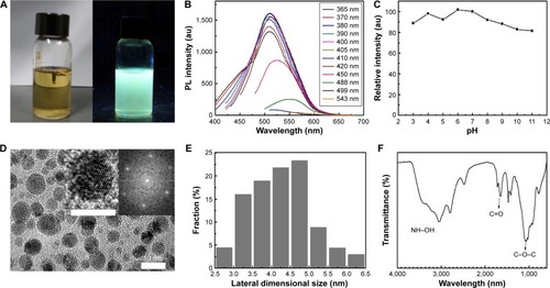
Figure 3 Viability studies of GQDs performed in DU-145 (A, B, and C) and PC-3 (D, E, and F) prostate cancer cell lines.
Notes: The quantification of viable cell number upon 24 h exposure to different concentrations of GQDs was performed with a CCK-8 kit (A and D). Specific staining of live (green) and dead (red) cells (B and E) was also performed after 24 h incubation with 200 µg/mL GQDs concentration. The corresponding images of the stained cells with transmitted light are also presented (C and F). The scale bars of figures B, C, E, and F represent 50 µm.
Abbreviations: GQD, graphene quantum dot; h, hours.
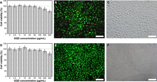
Figure 4 Optical properties of plain and functionalized GQDs.
Notes: (A) Ultraviolet-visible spectra of GQDs, free Dox, Dox-loaded GQDs (Dox-GQDs), and Dox-loaded RGD-modified GQDs (Dox-RGD-GQDs). (B) PL spectra of Dox-RGD-GQDs excited at 400 and 499 nm.
Abbreviations: Dox, doxorubicin; GQDs, graphene quantum dots; PL, photoluminescence; RGD, arginine-glycine-aspartic acid.
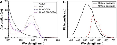
Figure 5 Kinetics of Dox release from Dox-RGD-GQDs at different pH values.
Notes: The values were obtained upon measuring the PL from the filtered solution (released Dox) and extrapolating the results to a calibration curve.
Abbreviations: Dox, doxorubicin; GQDs, graphene quantum dots; PL, photoluminescence; RGD, arginine-glycine-aspartic acid.
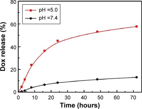
Figure 6 Viability tests of DU-145 (A), PC-3 (B), and MC3T3-E1 (C) cells after 24 hours incubation with free Dox, Dox-GQDs, and Dox-RGD-GQDs at different Dox concentrations.
Abbreviations: Dox, doxorubicin; GQDs, graphene quantum dots; RGD, arginine-glycine-aspartic acid.
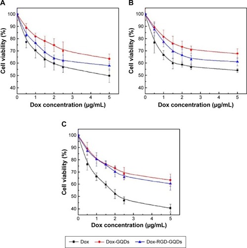
Figure 7 Confocal laser scanning microscope images of DU-145 (A, B, and C) and PC-3 (D, E, and F) cells treated with Dox-RGD-GQDs after incubation for 2, 8, and 16 hours at a Dox concentration of 2.5 µg/mL.
Notes: (a) GQDs excited by a 405 nm laser. (b) Dox excited by a 488 nm laser. (c) Merged images of GQDs and Dox. (d) Enlarged images of single cells from (c). (e) Fluorescence intensity of GQDs and Dox across the cell. Scale bar: 50 µm.
Abbreviations: Dox, doxorubicin; GQDs, graphene quantum dots; PL, photoluminescence; RGD, arginine-glycine-aspartic acid.
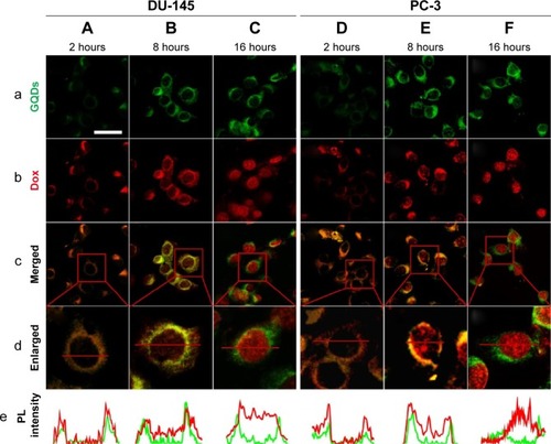
Figure 8 DU-145 (A, B, and C) and PC-3 (D, E, and F) cells were treated with Dox-RGD-GQDs for 2, 8, and 16 hours.
Notes: (a) Dox excited by a 488 nm laser. (b) Cell nuclei stained with DAPI excited by a 405 nm laser. (c) Merged images of Dox and DAPI. Scale bar: 50 µm.
Abbreviations: DAPI, 4′,6-diamidino-2-phenylindole; Dox, doxorubicin; GQDs, graphene quantum dots; RGD, arginine-glycine-aspartic acid.
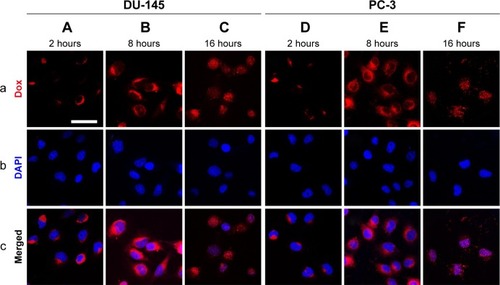
Figure S1 Characterization of GO.
Notes: (A) AFM images of GO sheets on mica surface. (B) Height profile of the AFM image. (C) Raman spectrum of GO.
Abbreviations: AFM, atomic force microscopy; GO, graphene oxide.
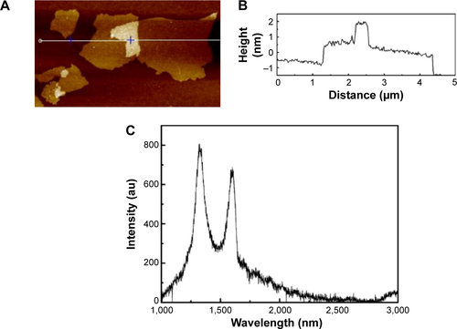
Figure S2 Transmission electron microscope images of GQDs (A, 20 nm; B, 10 nm).
Abbreviation: GQDs, graphene quantum dots.
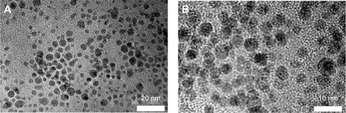
Figure S3 Confocal laser scanning microscope images of DU-145 (A and B) and PC-3 (C and D) cells after 24 hours incubation with GQDs at a concentration of 200 µg/mL.
Notes: Cells without GQDs were used as a control (B and D). GQDs were excited under a 405 nm laser. The results showed that under the same confocal laser scanning microscope conditions, cells incubated with GQDs emitted green fluorescence, while cells without GQDs showed no detectable fluorescence.
Abbreviation: GQDs, graphene quantum dots.
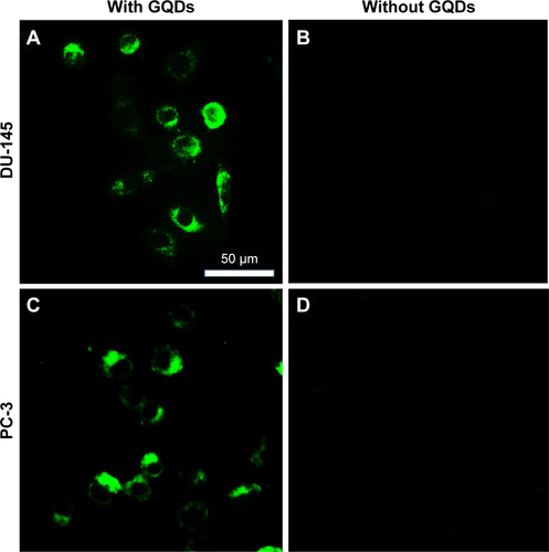
Figure S4 Ultraviolet-visible calibration curve of free Dox.
Abbreviation: Dox, doxorubicin.
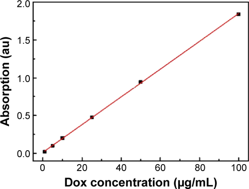
Figure S5 PL spectrum of Dox excited at wavelengths of 488 and 499 nm.
Abbreviations: Dox, doxorubicin; PL, photoluminescence.
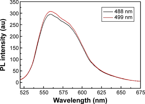
Figure S6 PL calibration curve of free Dox.
Abbreviations: Dox, doxorubicin; PL, photoluminescence.
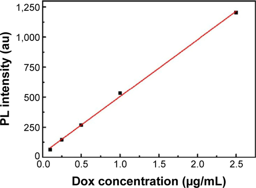
Figure S7 Confocal laser scanning microscope images of DU-145 (A, B, and C) and PC-3 (D, E, and F) cells after 16 hours incubation with GQDs.
Notes: GQDs are shown in the green channel, whereas the lysosomes of the cells are shown in the red channel. An overlay of both channels is also presented. Scale bar: 50 µm.
Abbreviation: GQDs, graphene quantum dots.
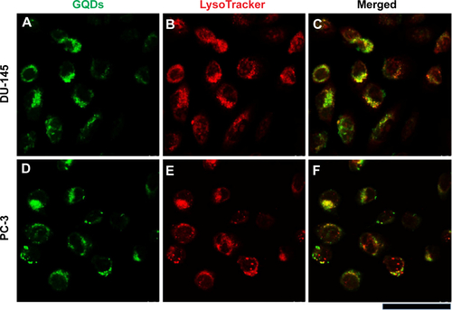
Figure S8 Confocal laser scanning microscope images of DU-145 (A and B) and PC-3 (C and D) cells treated with free Dox at a concentration of 2.5 µg/mL after 2 and 8 hours incubation.
Notes: (a) Blue regions are cell nuclei stained with DAPI excited by a 405 nm laser. (b) Red regions are Dox excited by a 488 nm laser. (c) Merged cells. Scale bar: 50 µm.
Abbreviations: DAPI, 4′,6-diamidino-2-phenylindole; Dox, doxorubicin.
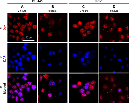
Table S1 Dox-loading efficiency onto Dox-GQDs and Dox-RGD-GQDs