Figures & data
Figure 1 (A) A top-view SEM image of TiO2 nanotubes (Ti/TiO2). (B) Effects of Ti/TiO2 surface vs cpTi on p38, ERK1/2, and JNK activation in the absence or presence of LPS in RAW 264.7 macrophages.
Notes: Cells were allowed to adhere on the surfaces for 24 hours and then incubated with 1 µg·mL−1 LPS for specified times. Phosphorylated proteins were detected by the ELISA technique as described in Materials and methods. The data are expressed as mean ± SD. *P<0.05; ***P<0.001.
Abbreviations: cpTi, commercial pure titanium; SEM, scanning electron microscope; JNK, c-Jun NH2-terminal kinase; LPS, lipopolysaccharide; ELISA, enzyme-linked immunosorbent assay; MAPK, mitogen-activated protein kinase; ERK, extracellular signal-regulated kinase; SD, standard deviation; vs, versus.
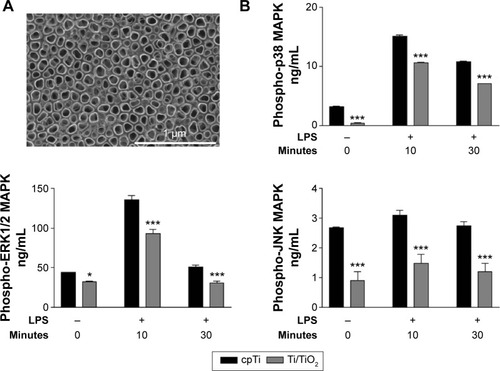
Figure 2 Effects of Ti/TiO2 nanotubular vs flat surface on IKKβ and IkB-α phosphorylation.
Notes: (A) RAW 264.7 cells were allowed to adhere on the substrates for 24 hours prior to stimulation with 1 µg·mL−1 LPS for specified times. The concentration of phospho-IKKβ in cell lysates was analyzed by the ELISA technique. The data are expressed as mean ± SD. **P<0.01; ***P<0.001. (B) Fluorescent immunodetection of p-IkB-α in untreated and LPS (1 µg·mL−1, for 10 minutes) stimulated macrophages using specific anti-p-IkB-α antibody (red). Nuclei were stained with DAPI (blue). Scale bar represents 20 µm.
Abbreviations: cpTi, commercial pure titanium; DAPI, 4′,6-diamidino-2-phenylindole; ELISA, enzyme-linked immunosorbent assay; LPS, lipopolysaccharide; OD, optical density; SD, standard deviation; vs, versus.
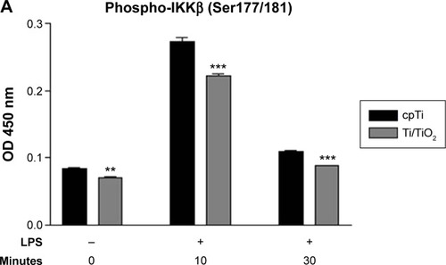
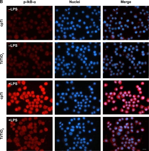
Figure 3 Effects of titania nanotubes vs cpTi on the nuclear translocation of NF-κB-p65.
Notes: (A) Fluorescent imaging of p65 in untreated and LPS-stimulated RAW 264.7 macrophages. Scale bar represents 10 µm. (B) Percentage of cells displaying nuclear accumulation of p65. Comparison was made between Ti/TiO2 and cpTi at 10 and 30 minutes poststimulation on six representative microscopic fields. The data are expressed as mean ± SD. ***P<0.001.
Abbreviations: cpTi, commercial pure titanium; LPS, lipopolysaccharide; NF-κB, nuclear factor kappa-light-chain-enhancer of activated B cells; SD, standard deviation; vs, versus.
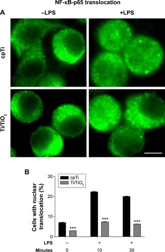
Figure 4 Effects of selective inhibitors of p38 (SB202190), ERK1/2 (U0126), and JNK (SP600125) on the production of TNF-α and MCP-1.
Notes: Cells were allowed to adhere on the samples for 18 hours, then were incubated with specific MAPK inhibitors for 1 hour and further treated with 1 µg·mL−1 LPS for 24 hours. The secreted pro-inflammatory factors were detected in cell culture media using ELISA assay. The data are expressed as mean ± SD. *P<0.05; **P<0.01; ***P<0.001.
Abbreviations: cpTi, commercial pure titanium; ELISA, enzyme-linked immunosorbent assay; JNK, c-Jun NH2-terminal kinase; LPS, lipopolysaccharide; MAPK, mitogen-activated protein kinase; MCP-1, monocyte chemotactic protein-1; TNF-α, tumor necrosis factor α; ERK, extracellular signal-regulated kinase; SD, standard deviation; vs, versus.
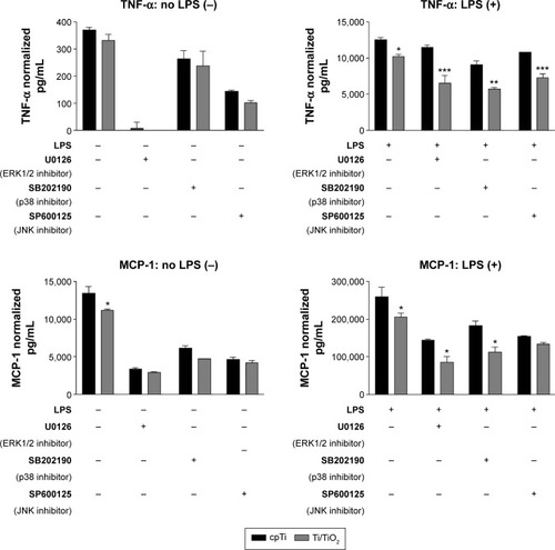
Figure 5 Nitrite concentrations in the cell culture media of LPS-stimulated macrophages under influence of selective inhibitors of p38 (SB202190), ERK1/2 (U0126), and JNK (SP600125).
Notes: Cells were allowed to adhere on the samples for 18 hours, then were incubated with selective MAPK inhibitors for 1 hour and further treated with 1 µg·mL−1 LPS for 24 hours. Nitrite accumulation in cell culture media was assessed by Griess reaction. The data are expressed as mean ± SD. *P<0.05; ***P<0.001.
Abbreviations: cpTi, commercial pure titanium; JNK, c-Jun NH2-terminal kinase; LPS, lipopolysaccharide; MAPK, mitogen-activated protein kinase; ERK, extracellular signal-regulated kinase; SD, standard deviation.
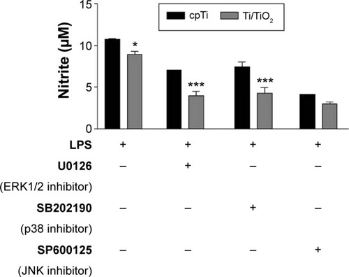
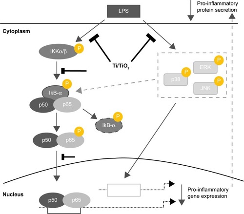
Figure 6 A schematic diagram showing the proposed mechanisms underlying the attenuation of the macrophage inflammatory response by TiO2 nanotubes.
Notes: Nanotubular TiO2 surface acts through significant suppression of LPS-induced NF-κB activation, an event correlated with its inhibitory effect on LPS-induced IkB kinase (IKK) activation, IkB-α phosphorylation (by IKKβ and, possibly, MAPKs), nuclear translocation of p65-NF-κB, and also through suppression of MAPK (ERK, p38, and JNK) phosphorylation. Solid arrows indicate the main inflammatory pathways activated by challenging the RAW 264.7 cells with LPS. The oblic dashed arrow indicates the potential involvement of MAPK in the regulation of NF-κB activation. The dotted arrows show the regulation of the pro-inflammatory gene expression. The right side vertical solid arrows denote the down-regulation of pro-inflammatory gene expression and protein secretion. The right side dashed arrow indicates the direct relationship between the level of protein secretion and that of gene expression. The blunt lines (┣) indicate the inhibition by Ti/TiO2 surfaces. The oval dashed line illustrates the degradation of the phosphorylated IkB-alpha. Yellow “P” symbol denotes the phosphorylated biomolecules.
Abbreviations: ERK, extracellular signal-regulated kinase; JNK, c-Jun NH2-terminal kinase; LPS, lipopolysaccharide; MAPK, mitogen-activated protein kinase; NF-κB, nuclear factor kappa-light-chain-enhancer of activated B cells.