Figures & data
Figure 1 Schematic representation of principle of photodynamic therapy: a photosensitizer is excited by an external light stimulus.
Note: This energy can be transferred to oxygen in tissue, which leads to the formation of reactive oxygen species, causing oxidation of cellular components (eg, proteins, lipids, and DNA) and finally cell death.
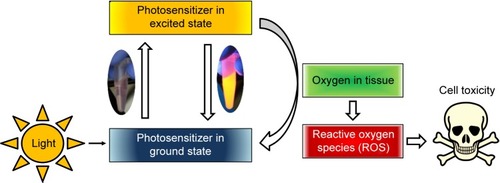
Table 1 Particle sizes (volume mean diameter) and ζ potential in water at pH 7
Figure 2 FT-IR spectra demonstrate the success of the functionalization of SPIONCMD.
Notes: The inset shows the introduction of peaks at 1,720 and 1,600 cm−1, which can be contributed to carboxyl groups (A). The spectra for SPIONCMD-Hyp shows changes in relative peak intensities, which can be attributed to the linkage of hypericin and glutaraldehyde; the inset shows magnification of the important wavenumber range (B).
Abbreviations: FT-IR, fourier transform infrared; SPIONCMD, functionalized dextran-coated SPIONs; SPION, superparamagnetic iron oxide nanoparticle; SPIONCMD-Hyp, hypericin linked to SPIONCMD; SPIONDEX, dextran-coated SPIONs; au, arbitrary unit.
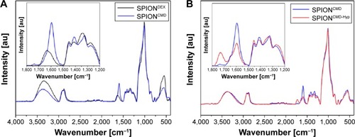
Figure 3 Exemplary HPLC ESI-MS chromatogram of hypericin (A). Linear calibration curve (R2=0.9993) for the determination of the particles’ hypericin concentration (B).
Abbreviations: HPLC ESI-MS, high-performance liquid chromatography combined with an electrospray-ionization mass spectrometer; m, minutes.
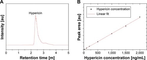
Figure 4 Overview TEM image of SPIONCMD (A) and SPIONCMD-Hyp (B) particles show agglomerates of SPIONs.
Abbreviations: TEM, transmission electron microscopy; SPIONCMD, functionalized dextran-coated SPIONs; SPION, superparamagnetic iron oxide nanoparticle; SPIONCMD-Hyp, hypericin linked to SPIONCMD.
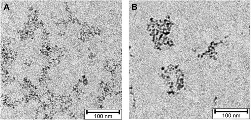
Figure 5 Magnetite particle distributions derived from measuring 200 particles of TEM images with the software ImageJ.
Notes: The sizes for SPIONCMD ranged from 3.0 to 9.0 nm and had a mean value of 4.5±1.0 nm (A). The sizes for SPIONCMD-Hyp ranged from 3.0 to 10.0 nm and had a mean value of 4.6±1.0 nm (B).
Abbreviations: TEM, transmission electron microscopy; SPIONCMD, functionalized dextran-coated SPIONs; SPION, superparamagnetic iron oxide nanoparticle; SPIONCMD-Hyp, hypericin linked to SPIONCMD.
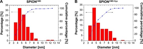
Figure 6 XRD pattern of SPIONCMD-Hyp exhibits typical peaks for the spinel structure of magnetite.
Note: The high noise is typical for nanocrystallites.
Abbreviations: XRD, X-ray diffraction; SPIONCMD, functionalized dextran-coated SPIONs; SPION, superparamagnetic iron oxide nanoparticle; SPIONCMD-Hyp, hypericin linked to SPIONCMD.
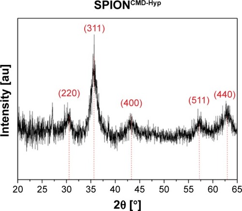
Figure 7 Cell death of Jurkat cells determined by DiI staining 24 hours (A) and 48 hours (B) after treatment with hypericin in different concentrations.
Notes: DiI-positive cells were considered viable, DiI-negative cells were considered dying/dead. Without illumination hypericin shows no toxicity, whereas with increased exposure time (0, 5, 10, and 15 minutes) as well as concentration (0, 0.1, 0.2, and 0.3 µg/mL) cell death can be observed. Figures show the mean values of triplicates with standard deviations. The statistical significance of comparisons with the untreated control was investigated using Student’s t-test in Excel (Microsoft Corporation, Redmond, WA, USA) (*P<0.05, **P<0.005, and ***P<0.0005).
Abbreviation: DiI, hexamethylindodicarbo-cyanine iodide.
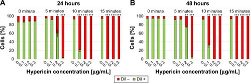
Figure 8 Cell death of Jurkat cells determined by DiI staining 24 hours (A) and 48 hours (B) after treatment with SPIONCMD, SPIONCMD-Hyp, SPIONCMD + hypericin not linked to the particles as well as hypericin alone at a hypericin concentration of 0.2 µg/mL.
Notes: DiI-positive cells were considered viable, DiI-negative cells were considered dying/dead. Without illumination all samples show no toxicity, whereas with increased exposure time (0, 5, 10, and 15 minutes) cell death can be observed. Figures show the mean values of triplicates with standard deviations. The statistical significance of comparisons with the untreated control was investigated using Student’s t-test in Excel (Microsoft Corporation, Redmond, WA, USA) (*P<0.05, **P<0.005, and ***P<0.0005).
Abbreviations: DiI, hexamethylindodicarbo-cyanine iodide; SPIONCMD, functionalized dextran-coated SPIONs; SPION, superparamagnetic iron oxide nanoparticle; SPIONCMD-Hyp, hypericin linked to SPIONCMD.
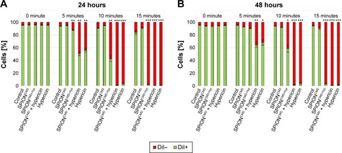
Figure 9 Determination of ROS generation (A) and corresponding cell death (B).
Notes: DCFH-DA-treated Jurkat cells after application of SPIONCMD, SPIONCMD-Hyp, SPIONCMD + hypericin not linked to the particles as well as hypericin alone with a drug concentration of 0.2 µg/mL and exposure time of 1 minute. Cell viability was determined by morphological cell analysis. After incubation for 5 minutes the hypericin samples showed ROS generation and correspondent cell death, whereas hypericin-bound nanoparticles without illumination did not generate ROS. Figures present the mean value of quadruplicates with standard deviations. The statistical significance of comparisons with the untreated control was investigated using Student’s t-test in Excel (Microsoft Corporation, Redmond, WA, USA) (*P<0.05, **P<0.005, and ***P<0.0005).
Abbreviations: DCFH-DA, 2′,7′-dichlorofluorescein diacetate; SPIONCMD, functionalized dextran-coated SPIONs; SPION, superparamagnetic iron oxide nanoparticle; SPIONCMD-Hyp, hypericin linked to SPIONCMD; ROS, reactive oxygen species; au, arbitrary unit.
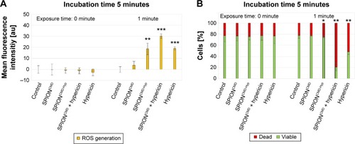
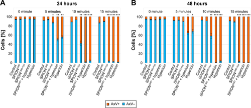
Figure S1 Cell death of Jurkat cells determined by AxV staining 24 hours (A) and 48 hours (B) after treatment with SPIONCMD, SPIONCMD-Hyp, and SPIONCMD + hypericin not linked to the particles as well as hypericin alone at a hypericin concentration of 0.2 µg/mL.
Notes: AxV-negative cells were considered viable, AxV-positive cells were considered dying/dead. Without illumination all samples show no toxicity, whereas with increased exposure time (0, 5, 10, and 15 minutes) cell death can be observed. Figure shows the mean values of triplicates with standard deviations. The statistical significance of comparisons with the untreated control was investigated using Student’s t-test in Excel (Microsoft Corporation, Redmond, WA, USA) (*P<0.05, **P<0.005, and ***P<0.0005).
Abbreviations: AxV, Annexin V; SPIONCMD, functionalized dextran-coated SPIONs; SPION, superparamagnetic iron oxide nanoparticle; SPIONCMD-Hyp, hypericin linked to SPIONCMD.