Figures & data
Figure 1 A brief workflow of the preparation of HPEI nanoparticles.
Abbreviations: HPEI, heparin–polyethyleneimine; PEI, polyethylenimine.
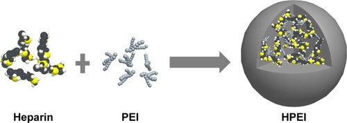
Figure 2 Characterization of HPEI nanoparticles.
Notes: (A) Size distribution spectrum of HPEI nanoparticles. (B) Zeta potential spectrum of HPEI nanoparticles. (C) Transmission electron microscopic image of HPEI nanoparticles. (D) The DNA-binding ability of HPEI nanoparticles determined by gel retardation assay. All data were representative of three independent experiments.
Abbreviations: HPEI, heparin–polyethyleneimine; DNA, deoxy ribonucleic acid; N:P, ratio of nitrogen atoms to phosphate group.
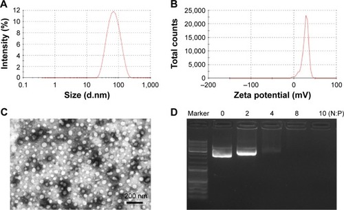
Figure 3 Mass spectrometry qualitatively determined the degradation of HPEI nanoparticles postintraperitoneal injection.
Notes: (A) Mass spectrum of standard PEI 2K. (B) Mass spectrum of the urine extracted from a HPEI-treated rat. (C) Mass spectrum of urine extracted from PEI 2K-treated rats. (D) Mass spectrum of urine extracted from PEI 25K-treated rats.
Abbreviations: HPEI, heparin–polyethyleneimine; PEI 2K, polyethyleneimine (molecular weight 2,000); PEI 25K, polyethyleneimine (molecular weight 25,000).
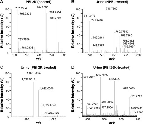
Figure 4 Representative ex vivo fluorescence imaging of tissues (heart, liver, spleen, lung, and kidney) harvested from the euthanized nude mice at 1, 2, 4, 6, 8, 24, and 30 hours post FITC–HPEI injection.
Abbreviations: FITC, fluorescein isothiocyanate; HPEI, heparin–polyethyleneimine; NS, normal saline; h, hours; Min, minimum; Max, maximum.
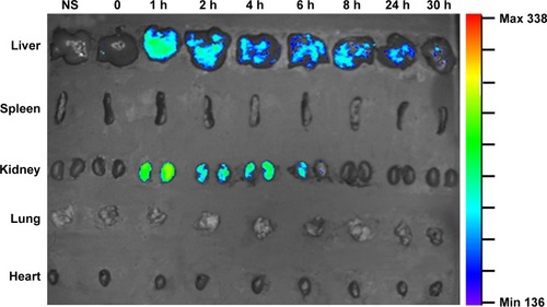
Figure 5 Cytotoxicity and transfection efficiency of HPEI nanoparticles.
Notes: (A) SKOV3 cells were treated with PEI 25K or HPEI in various concentrations for 48 hours, cell viability was measured by MTT assay. (B) The transfection efficiency of PEI 2K, HPEI, or PEI 25K was determined by flow cytometry, respectively. pGFP was used as a report gene. (C) Fluorescent image of SKOV3 cells transfected by pGFP–HPEI. Scale bar, 100 μm. (D) Fluorescent image of SKOV3 cells transfected by pGFP–PEI 25K. Scale bar, 100 μm. All data were representative of three independent experiments.
Abbreviations: HPEI, heparin–polyethyleneimine; PEI 2K, polyethyleneimine (molecular weight 2,000); PEI 25K, polyethyleneimine (molecular weight 25,000).
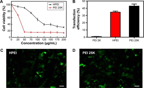
Figure 6 The plasmid map of pVITRO2–hs-T34A.
Abbreviations: hs, human survivin; bp, base pair.
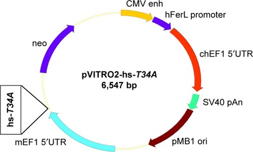
Figure 7 Anticancer effect of HPEI/hs-T34A complexes in vitro.
Notes: (A) and (B) HPEI/hs-T34A complexes were transfected in SKOV3 cells for 48 hours, the expression of survivin was examined by Western blot assay (A) and real time RT-PCR (B). (C) SKOV3 cells were treated with NS, HPEI alone (50 μg), HPEI/EP (50 μg/5 μg), or HPEI/hs-T34A complexes (50 μg/5 μg) for 48 hours. Cells were then stained with propidium iodide to evaluate apoptotic ratio by flow cytometric analysis. All data were representative of three independent experiments.
Abbreviations: HPEI, heparin–polyethyleneimine; hs, human survivin; RT-PCR, reverse-transcription polymerase chain reaction; EP, empty vector plasmid; NS, normal saline.
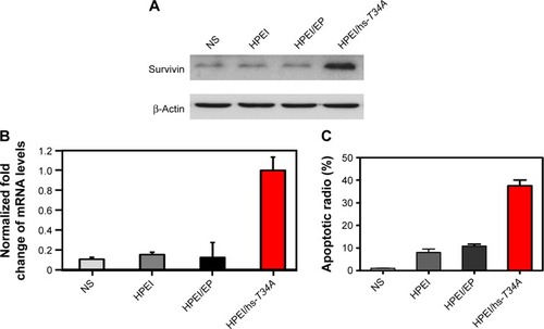
Figure 8 HPEI/hs-T34A complexes inhibited the growth of ovarian tumor in vivo.
Abbreviations: HPEI, heparin–polyethyleneimine; PEI 25K, polyethyleneimine (molecular weight 25,000); EP, empty vector plasmid; hs, human survivin.
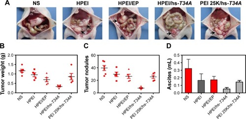
Figure 9 The expression of survivin in tumor tissues.
Notes: Western blot analysis (A) and real time RT-PCR (B) of ovarian tumor tissues in normal saline (NS), HPEI alone (50 μg), HPEI/EP, HPEI/hs-T34A, or PEI 25K/hs-T34A group, respectively.
Abbreviations: HPEI, heparin–polyethyleneimine; PEI 25K, polyethyleneimine (molecular weight 25,000); EP, empty vector plasmid; hs, human survivin; RT-PCR, reverse transcription polymerase chain reaction.
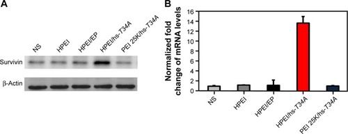
Figure 10 The effect of HPEI/hs-T34A complexes on apoptosis and angiogenesis of ovarian tumor cells.
Notes: (A) TUNEL assay was carried out to examine the apoptotic cells of ovarian tumor in NS, HPEI alone, HPEI/EP, or HPEI/hs-T34A complexes group, respectively. Scale bar, 50 μm. (B) CD31 staining was performed to assess angiogenesis of ovarian tumor in NS, HPEI alone, HPEI/EP, or HPEI/hs-T34A complexes group, respectively. Scale bar, 50 μm. The arrows show the CD31 positive vascular tissue.
Abbreviations: HPEI, heparin–polyethyleneimine; TUNEL, terminal deoxynucleotidyl transferase dUTP nick end labeling; EP, empty vector plasmid; NS, normal saline; CD31, clusters of differentiation 31; hs, human survivin.
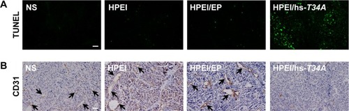
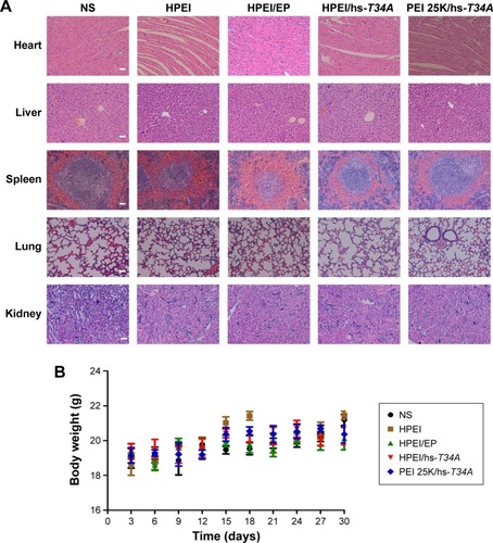
Figure 11 Safety and toxicity evaluation of HPEI/hs-T34A formulation.
Notes: (A) The heart, liver, spleen, lung, and kidney in NS, HPEI alone (50 μg), HPEI/EP, HPEI/hs-T34A, or PEI 25K/hs-T34A group were collected and conducted with HE staining, respectively. Scale bar, 50 μm. (B). Body weight changes in NS, HPEI alone (50 μg), HPEI/EP, HPEI/hs-T34A, or PEI 25K/hs-T34A group, respectively.
Abbreviations: HPEI, heparin–polyethyleneimine; PEI 25K, polyethyleneimine (molecular weight 25,000); HPEI/EP, heparin–polyethyleneimine/empty vector plasmid; NS, normal saline; hs, human survivin.