Figures & data
Figure 1 (A and B) High-resolution scanning electron microscopy of the bare MNPs and the thawed scaffold with MNPs; (C and D) scanning electron microscopy of the nanofiber scaffold with and without MNPs.
Notes: (A) MNPs (magnification ×65,000); (B) thawed PCL-MNPs (magnification ×20,000); (C) PCL-MNPs (magnification ×7,500); (D) PCL scaffold (magnification ×12,000).
Abbreviations: MNPs, magnetic nanoparticles; PCL-MNPs, poly-ε-caprolactone scaffold with magnetic nanoparticles; PCL, poly-ε-caprolactone.
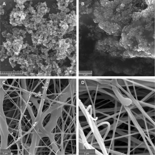
Figure 2 (A and B) Powder X-ray diffractogram and Mössbauer spectrum of the MNPs, both measured at room temperature. Notes: (A) X-ray diffractogram of the MNPs; (B) Mössbauer spectrum of the MNPs.
Abbreviation: MNPs, magnetic nanoparticles.
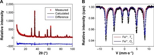
Figure 3 (A and B) Comparison of the magnetic characterization of the bare MNPs and the scaffold with MNPs.
Notes: (A) Magnetization isotherms recorded at low temperature and at room temperature, respectively; (B) the temperature dependence of the ZFC and FC magnetization for bare MNPs and the ZFC curve for the scaffold PCL-MNPs. The kink at approximately 350 K on the ZFC curve of the scaffold sample corresponds to melting of the fibers.
Abbreviations: MNPs, magnetic nanoparticles; PCL-MNPs, poly-ε-caprolactone scaffold with magnetic nanoparticles; ZFC, zero-field-cooled; FC, field-cooled.
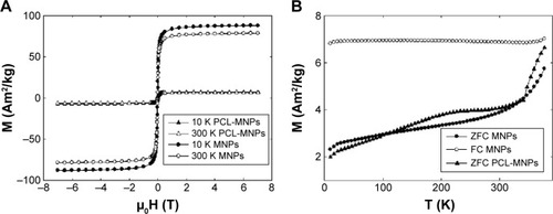
Figure 4 (A and B) Detail of the hysteresis loops recorded at low temperature and at room temperature, respectively.
Notes: (A) Hysteresis loop for the bare MNPs; (B) hysteresis loop for the PCL-MNPs. A moderate increase in the coercivity of the scaffold samples can be observed at 10 K.
Abbreviations: MNPs, magnetic nanoparticles; PCL-MNPs, poly-ε-caprolactone scaffold with magnetic nanoparticles.
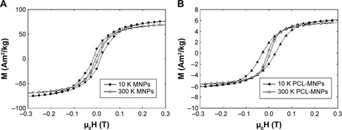
Figure 5 Metabolic activity of the MSCs measured by MTS assay.
Notes: Statistical analysis: *P<0.05; **P<0.001.
Abbreviations: MSCs, mesenchymal stem cells; MTS, 3-(4,5-dimethylthiazol-2-yl)-5-(3-carboxymethoxyphenyl)-2-(4-sulfophenyl)-2H-tetrazolium; PCL, poly-ε-caprolactone scaffold; PCL-MNPs, poly-ε-caprolactone scaffold with magnetic nanoparticles.
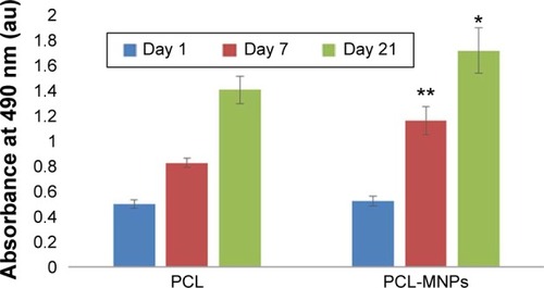
Figure 6 Proliferation of the MSCs determined using PicoGreen assay.
Notes: Statistical analysis: *P<0.05; **P<0.001.
Abbreviations: MSCs, mesenchymal stem cells; PCL, poly-ε-caprolactone scaffold; PCL-MNPs, poly-ε-caprolactone scaffold with magnetic nanoparticles.
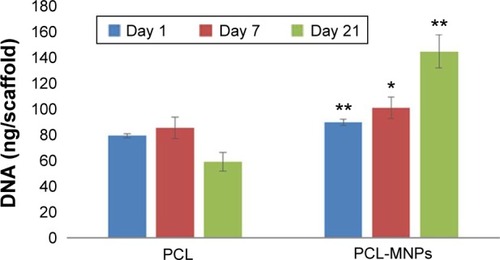
Figure 7 (A–F) Fluorescent staining of the MSCs cultured on PCL and PCL-MNPs, respectively. Phalloidin-rhodamine was used to stain cytoplasm (red), DNA was stained by Hoechst 33342 (blue).
Notes: (A) MSCs on PCL scaffold on day 1; (B) MSCs on PCL-MNPs scaffold on day 1; (C) MSCs on PCL scaffold on day 7; (D) MSCs on PCL-MNPs scaffold on day 7; (E) MSCs on PCL scaffold on day 21; (F) MSCs on PCL-MNPs scaffold on day 21.
Abbreviations: MSCs, mesenchymal stem cells; PCL, poly-ε-caprolactone scaffold; PCL-MNPs, poly-ε-caprolactone scaffold with magnetic nanoparticles.
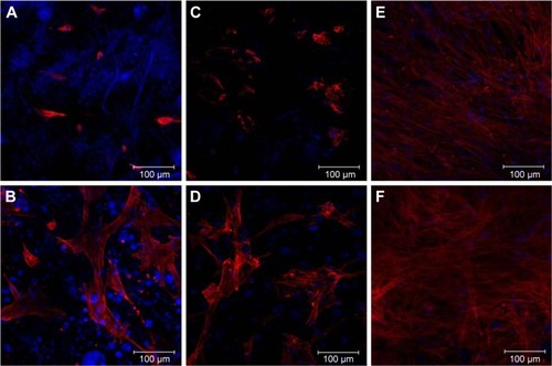
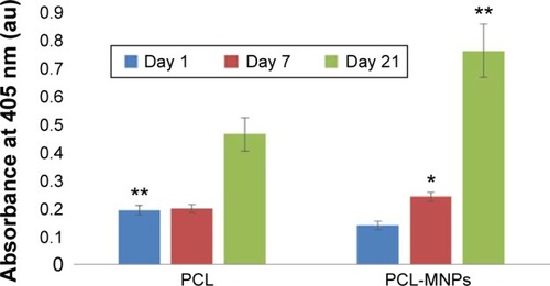
Figure 8 Alkaline phosphatase activity of the MSCs.
Notes: Statistical analysis: *P<0.05; **P<0.001.
Abbreviations: MSCs, mesenchymal stem cells; PCL, poly-ε-caprolactone scaffold; PCL-MNPs, poly-ε-caprolactone scaffold with magnetic nanoparticles.