Figures & data
Figure 1 SPION uptake in MSCs and their corresponding magnetic heating characteristics.
Notes: (A) SQUID measurements of the concentration of iron oxide per mesenchymal stem cell (MSC) after overnight incubation of 0.5 mg/mL of FluidMAG-CT (citric acid – 50 nm), FluidMAG-CMX (carboxylmethyldextran), FluidMAG-DX (dextran), FluidMAG-D (starch), and Ferucarbotran (carboxydextran). (B) MSCs take up SPIONs without affecting their phenotype; (i) MSCs in culture; (ii) Perl’s Prussian blue staining of MSCs after overnight culture with Ferucarbotran nanoparticles; (iii) differentiation to osteoblasts, Alizarin Red S staining; (iv) differentiation to adipocytes, Oil Red O staining. (C) Representative fiber-optic thermometry measurements of Ferucarbotran and FluidMAG-CT (50 nm- and 100 nm-sized SPIO particles) at 1 mg/mL. (D) Representative fiber-optic thermometry measurements of cell heating of a 5×10⁵ MSC pellet after overnight incubation of 0.5 mg/mL of FluidMAG-CT 50 nm or Ferucarbotran (representative of three experiments).
Abbreviations: SQUID, superconducting quantum interference device; SPION, superparamagnetic iron oxide nanoparticle; sec, seconds.
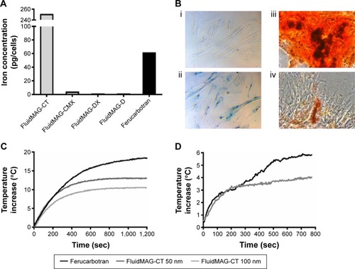
Figure 2 Murine model of OVCAR-3 tumor growth, with and without magnetic heating.
Notes: (A) Experimental plan of MRI and AMF heating. (B) T2 weighted MR images of a heated and nonheated OVCAR-3 tumor coinjected with Ferucarbotran-labeled MSCs, at days 14 (prior to heating), 18, and 22 (postheating) (white arrow indicates signal recovery). (C) Thermocamera image of a heated tumor (T) on the flank of a nude mouse (F) with C indicating the MACH coil and P the fiber-optic thermometry probe. (D) Fiber-optic thermometry measurements of a heated tumor over days 15, 17, 19, and 21 and a representative nonheated tumor and core body temperature. (E) Post-innoculation OVCAR-3 tumor growth for both heated and nonheated tumors.
Abbreviations: MRI, magnetic resonance imaging; AMF, alternating magnetic field; MR, magnetic resonance; MACH, magnetic alternating current hyperthermia; sec, seconds; MSCs, mesenchymal stem cells.
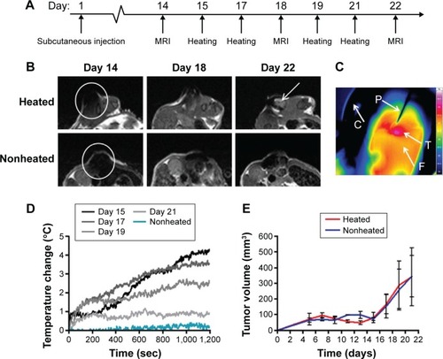
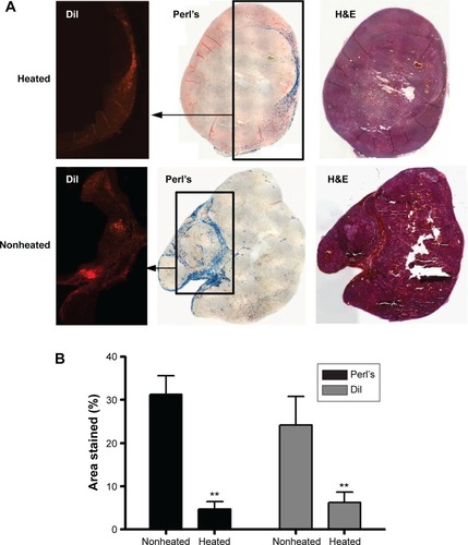
Figure 3 Immunohistochemistry data from a human OVCAR-3 tumor model, with and without magnetic heating.
Notes: (A) Sections from a representative tumor stained with DiI cell tracker dye, Perl’s Prussian blue iron stain, and H&E on day 22, derived from a coinjection, on day 1, of Ferucarbotran-labeled MSCs and OVCAR-3 tumor cells that were subsequently either heated or nonheated. (B) Percentage stained areas of representative slices throughout both heated and nonheated tumors for both Perl’s and DiI staining (error bars are SEM and **P<0.01).
Abbreviations: H&E, hematoxylin and eosin; MSCs, mesenchymal stem cells; SEM, standard error of the mean.