Figures & data
Figure 1 Schematic illustration of PET artificial ligament and collagen/simvastatin microspheres coating on PET in the artificial ligament anterior cruciate ligament (ACL) reconstruction.
Abbreviations: PET, polyethylene terephthalate; SIM/COL/PET, collagen and simvastatin microspheres collagen coating on polyethylene terephthalate fibers.
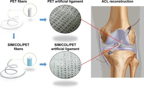
Figure 2 Digital images of the grafts and ACL reconstruction operation.
Notes: (A) Gross observation of PET, COL/PET, and SIM/COL/PET scaffolds. (B, a) Macroscopic view of rabbit knee joint (the arrow points to ACL rupture). (B, b) Macroscopic view of ACL reconstruction (the arrow points to implant).
Abbreviations: PET, polyethylene terephthalate; COL/PET, collagen coating on polyethylene terephthalate scaffolds; SIM/COL/PET, collagen and simvastatin microspheres coating on polyethylene terephthalate scaffolds; ACL, anterior cruciate ligament.
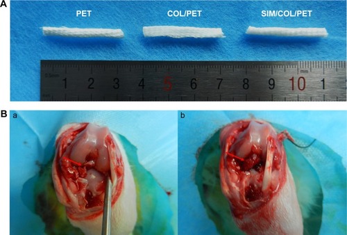
Figure 3 Scanning electron microscopic images of morphologic changes and cell adhesion in different fibers at 1 day and 4 days (100× magnification).
Notes: Micromorphology: original PET fibers (A), COL/PET (D), SIM/COL/PET (G); BMSCs adhesion: PET (B, C), COL/PET (E, F), and SIM/COL/PET (H, I). Insets show magnified sections of A-I (10× magnification).
Abbreviations: PET, polyethylene terephthalate; COL/PET, collagen coating on polyethylene terephthalate scaffolds; SIM/COL/PET, collagen and simvastatin microspheres coating on polyethylene terephthalate scaffolds; BMSCs, bone marrow stromal cells.
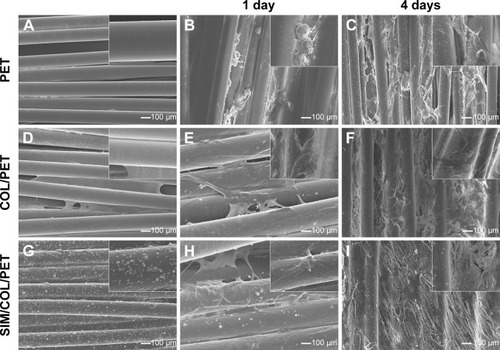
Figure 4 Characterization of the PET coating.
Notes: (A) Morphology characterization of simvastatin/polycaprolactone (SIM/PCL) microspheres. Inset shows the morphology of a single microsphere. (B) Simvastatin release profiles for SIM/PCL microspheres. (C) Water contact angle images of PET (85.25°±8.19°), COL/PET (33.68°±4.94°), and SIM/COL/PET (34.11°±6.12°).
Abbreviations: PET, polyethylene terephthalate; COL/PET, collagen coating on polyethylene terephthalate scaffolds; SIM/COL/PET, collagen and simvastatin microspheres coating on polyethylene terephthalate scaffolds; SIM, simvastatin.
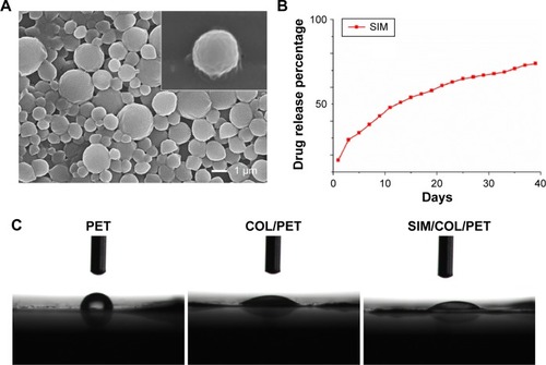
Figure 5 Measurement of BMSC proliferation and adhesion among the PET group, COL/PET group, and SIM/COL/PET group.
Notes: (A) Cell adhesion ability on different samples and (B) BMSC viability in the scaffolds was detected with cell counting kit-8 (CCK-8). *P<0.05 compared with control; #P<0.05 between treatment groups.
Abbreviations: PET, polyethylene terephthalate; COL/PET, collagen coating on polyethylene terephthalate scaffolds; SIM/COL/PET, collagen and simvastatin microspheres coating on polyethylene terephthalate scaffolds; BMSC, bone marrow stromal cell; OD, optical density.
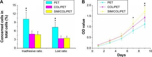
Figure 6 The results of the mineralization of BMSCs seeding on PET samples.
Notes: (A) The ALP activity of BMSCs seeded on PET, COL/PET, and SIM/COL/PET scaffolds for 7 days and 14 days. (B) Alizarin red staining after BMSCs seeded on these scaffolds for 21 days. (C) The ALP quantitative analysis for 7 days and 14 days. (D) Alizarin red staining quantitative analysis (415 nm) for 21 days. The osteogenic-related gene expressions (OC, RUNX-2, BMP-2, VEGF) in different groups at days 1 (E) and 3 (F). *P<0.05; **P<0.01 compared with control; #P<0.05 between treatment groups.
Abbreviations: PET, polyethylene terephthalate; COL/PET, collagen coating on polyethylene terephthalate scaffolds; SIM/COL/PET, collagen and simvastatin microspheres coating on polyethylene terephthalate scaffolds; BMSCs, bone marrow stromal cells; ALP, alkaline phosphatase; BMP-2, bone morphogenetic protein-2; RUNX-2, runt-related transcription factor 2; OC, osteocalcin; VEGF, vascular endothelial growth factor; OD, optical density.
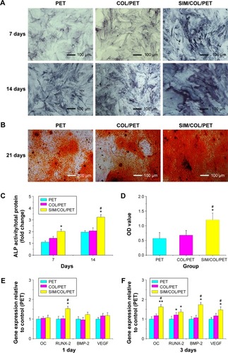
Figure 7 Histological and immunohistological assessments.
Notes: (A) Hematoxylin and eosin staining evaluation of the graft–bone interface at 4 weeks and 8 weeks after surgery (magnification, ×200). The black arrow shows the tendon–bone interface. (B) Immunohistochemical staining of the osteogenic and angiogenesis marker, VEGF, at 4 weeks and 8 weeks after surgery (magnification, ×100). The brown precipitate represented stained VEGF (red arrows).
Abbreviations: G, artificial ligament graft; IF, interface; VEGF, vascular endothelial growth factor; PET, polyethylene terephthalate; COL/PET, collagen coating on polyethylene terephthalate scaffolds; SIM/COL/PET, collagen and simvastatin microspheres coating on polyethylene terephthalate scaffolds.
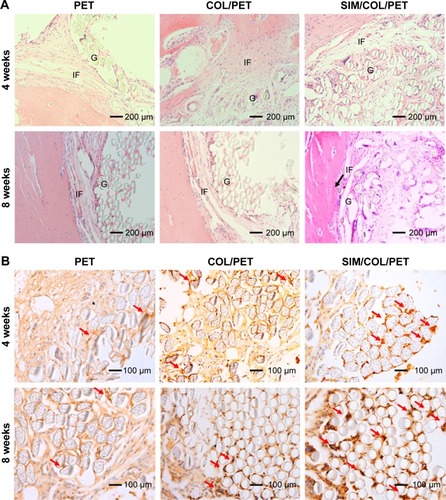
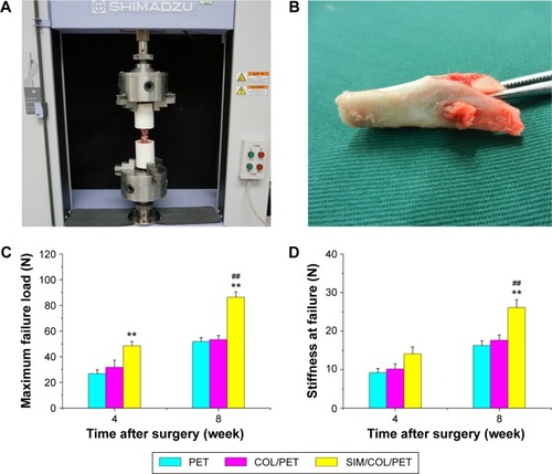
Figure 8 Mechanical examinations for graft–bone healing in a rabbit model at each time point after surgery.
Notes: (A) Digital camera image of biomechanical test experiment of implanted graft. (B) PET graft was in the tibial bone tunnel. (C) Comparison of maximal failure load among the PET group, COL/PET group, and SIM/COL/PET group. (D) Comparison of stiffness at failure among the PET group, COL/PET group, and SIM/COL/PET group. **P<0.01 compared with control; ##P<0.01 between treatment groups.
Abbreviations: PET, polyethylene terephthalate; COL/PET, collagen coating on polyethylene terephthalate scaffolds; SIM/COL/PET, collagen and simvastatin microspheres coating on polyethylene terephthalate scaffolds.