Figures & data
Table 1 Effect of gold and PZQ treatment on worms burden and egg count of schistosome infected mice
Figure 1 Typical TEM and corresponding HR-TEM images of synthesized AuNPs.
Notes: (A) A low-magnification image of spherical AuNPs (~10–15 nm). (B) An HR-TEM image of the difference between two lattice fringes, which iŝ0.233 nm.
Abbreviations: TEM, transmission electron microscopy; HR-TEM, high-resolution transmission electron microscopy; AuNPs, gold nanoparticles.
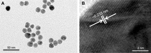
Figure 2 Egg density-induced changes in the livers of mice infected with Schistosoma mansoni and treated with AuNPs.
Notes: Values are mean ± SD (n=8). *Significant change at P≤0.05.
Abbreviations: AuNPs, gold nanoparticles; SD, standard deviation; PZQ, praziquantel.
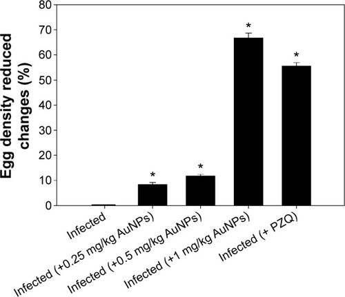
Table 2 AuNPs induced alterations in the hepatic tissue of mice infected with Schistosoma mansoni
Figure 3 Histological changes in hepatic tissue of noninfected mice, untreated mice, and mice treated and infected with Schistosoma mansoni on day 56 postinfection.
Notes: (A) Noninfected liver with normal architecture. (B) Hepatic tissue of mice in the S. mansoni-infected group showing a severe inflammatory response in the liver indicated by inflammatory cellular infiltration, cytoplasmic vacuolation, degeneration of hepatocytes, dilated hepatic sinusoids dilated, and more Kupffer cells. (C–E) Hepatic tissue of mice in the S. mansoni-infected group treated with 0.25, 0.5, and 1 mg/kg AuNPs, respectively, showing reduced tissue damage, and (F) the liver of mice in the infected group treated with PZQ showing fewer lesions. Arrows indicate the inflammatory cellular infiltraion around the granuloma and arrow heads indicates enclosed eggs. Sections were stained with hematoxylin and eosin; scale bar =25 μm.
Abbreviations: AuNPs, gold nanoparticles; PZQ, praziquantel.
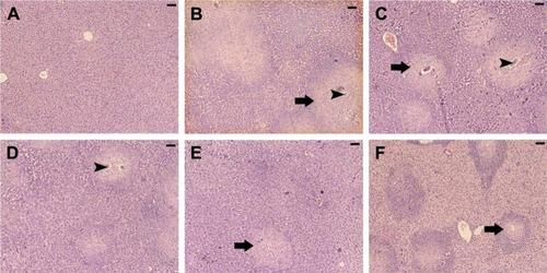
Figure 4 Reduction in granuloma size in the livers of mice infected with Schistosoma mansoni and treated with AuNPs.
Notes: Values are mean ± SD (n=8). *Significant change at P≤0.05.
Abbreviations: AuNPs, gold nanoparticles; SD, standard deviation; PZQ, praziquantel.
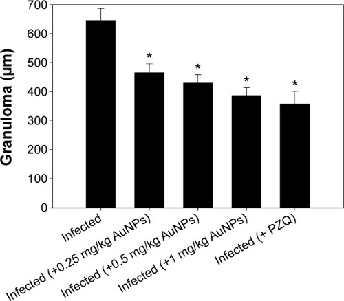
Table 3 AuNPs induced changes in the level of hepatic GSH, MDA, and NO of mice infected with Schistosoma mansoni
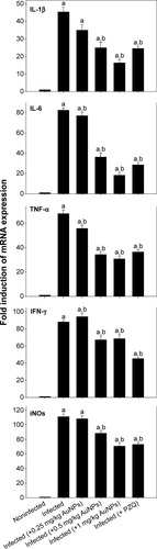
Figure 5 AuNPs induced changes in gene expression of mice livers infected with Schistosoma mansoni.
Notes: Expression of IL-1β, IL-6, TNF-α, IFN-γ, and iNOS in liver tissues was analyzed by quantitative RT-PCR in noninfected mice and S. mansoni-infected mice on day 56 postinfection with and without AuNPs treatment. Relative expression is given as fold increase in comparison with noninfected control mice. Values are mean ± SD. aSignificant against the noninfected (AuNPs) group at P≤0.05. bSignificant against the infected (AuNPs) group at P≤0.05.
Abbreviations: AuNPs, gold nanoparticles; IL, interleukin; TNF-α, tumor necrosis factor-α; IFN-γ, interferon-γ; iNOS, inducible nitric oxide synthase; RT-PCR, real-time polymerase chain reaction; SD, standard deviation; PZQ, praziquantel.