Figures & data
Table 1 Primer sequences and conditions for RT-PCR analysis
Figure 1 TEM image of PLL-BNNT dispersion: both single nanotubes (red arrows) and small aggregates of nanotubes are visible.
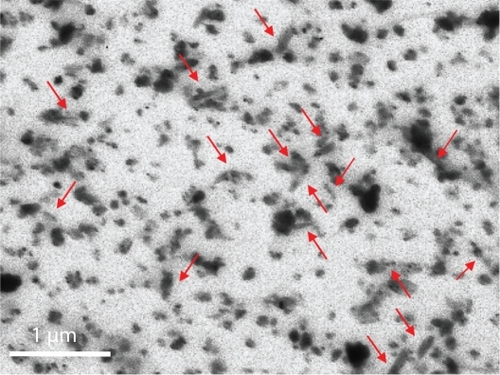
Figure 2 Confocal analysis of BNNT internalization a) and investigation of its mechanism: BNNT uptake b) and its inhibition after treatment with sodium azide c).
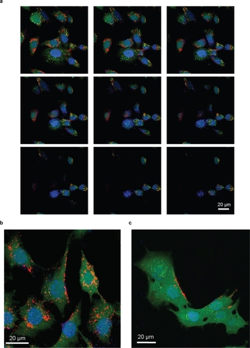
Figure 3 TEM imaging of C2C12 cells incubated for 12 h with (a) and without (b) a 10 μg/mL BNNT modified medium. The TEM micrograph shows C2C12 with vesicles containing BNNT (arrows) in the treated sample. Magnification 7000×.
Abbreviations: BNNT, boron nitride nanotubes; M, mitochondria; N, nuclei; TEM, transmission electron microscopy.
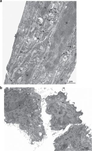
Figure 4 MTT assay results after 24, 48, and 72 h of incubation of C2C12 cells with 0, 5, 10, and 15 μg/mL of PLL-BNNTs, and with 3 μg/mL of PLL (n = 6).
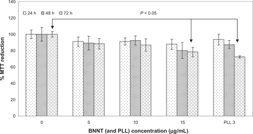
Figure 5 LIVE/DEAD® viability/cytotoxicity assay and early apoptotic detection (annexin V-FITC/PI assay) performed for different incubation times and concentrations.
Note: Scale bar: 100 μm.
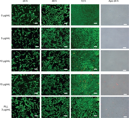
Figure 6 Total protein content normalized by ds-DNA content in C2C12 cell differentiated in presence of 0, 5, and 10 μg/mL of PLL-BNNTs (n = 3, *P < 0.05).
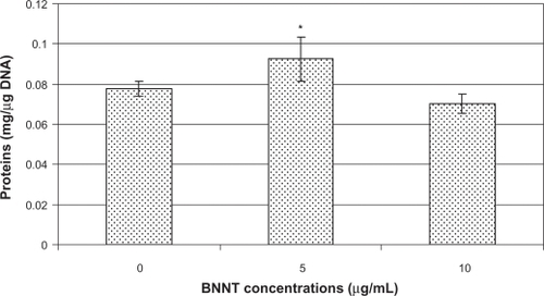
Figure 7 Light microscopy on H&E-stained cell samples showing myotube formation: differentiated C2C12 cultured with 0 (a), 5 (b), and 10 (c) μg/mL of PLL-BNNTs; fusion index evaluated for the three cultures (d).
Abbreviations: BNNTs, boron nitride nanotubes; H&E, hematoxylin–eosin; PLL, poly-l-lysine.
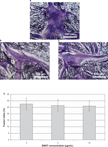
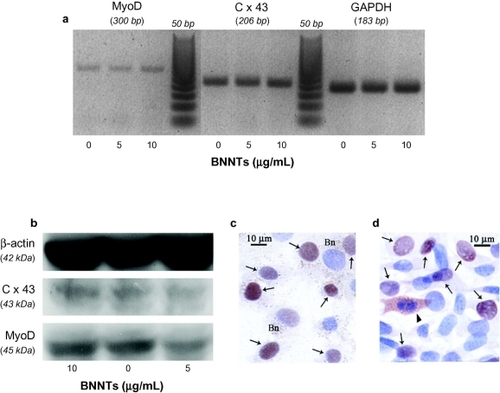
Figure 8 RT-PCR results for MyoD and Cx43 expression in C2C12 cells differentiated in presence of 0, 5, and 10 μg/ml of PLL-BNNTs; GAPDH was used as internal standard (a) Western blot results for MyoD and Cx43 production in the same cultures; β-actin was used as internal standard (b) Immunocytochemical analysis for MyoD expression on BNNT-treated cell samples (c) and controls without BNNTs (d) “Bn” indicates visible dotting in the cytoplasm due to BNNTs, the arrows indicate MyoD positive cells at nuclear level, while the arrowhead indicates MyoD positive cells at cytoplasm level.
Abbreviations: BNNTs, boron nitride nanotubes; H&E, hematoxylin–eosin; PLL, poly-l-lysine; RT-PCR, reverse transcription – polymerase chain reaction.