Figures & data
Figure 1 SEM images of nonmodified PLA membranes.
Notes: Quanta 450 scanning electron microscope FEI, magnification 2,000× (A), magnification 10,000× (B). Morphological parameters of PLA membranes (C). Mean ± SD from 12 SEM images (1,748 measurements in total).
Abbreviations: SEM, scanning electron microscopy; PLA, polylactide; SD, standard deviation.

Figure 2 Morphology of the fibrin nanocoating (F) on PLA membranes.
Notes: The fibrin nanocoating stained with primary and secondary antibody. Arrows show the thin fibrous mesh of fibrin at the top of the membrane. The nonmodified membranes (PLA) were used as a control material to show nonspecific binding of primary or secondary antibody. Leica TCS SPE DM2500 confocal microscope, obj 40×/1.15 NA oil.
Abbreviations: PLA, polylactide; obj, objective; NA, numerical aperture; PLA F, fibrin nanocoating on PLA.
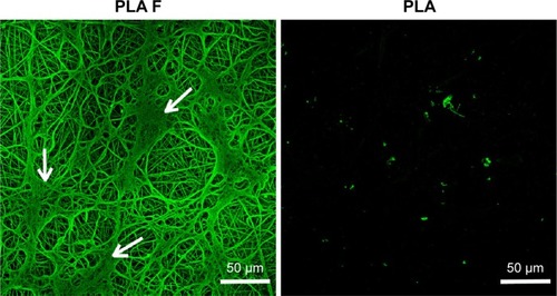
Figure 3 Morphology of the fibrin nanocoating (green) on PLA membranes in six cell culture intervals incubated without cells at 37°C, 5% CO2 (A), incubated with human dermal fibroblasts (B – only fibrin nanocoating, C – only cells, D – fibrin nanocoating with cells). Cells on nonmodified PLA membranes (E).
Notes: Cells cultivated in the standard cell culture medium. The fibrin nanocoating was stained by immunofluorescence. The cells were stained with phalloidin conjugated with TRITC and Hoechst #33258. Leica TCS SPE DM2500 confocal microscope, obj 40×/1.15 NA oil.
Abbreviations: PLA, polylactide; obj, objective; NA, numerical aperture; PLA F, fibrin nanocoating on PLA.
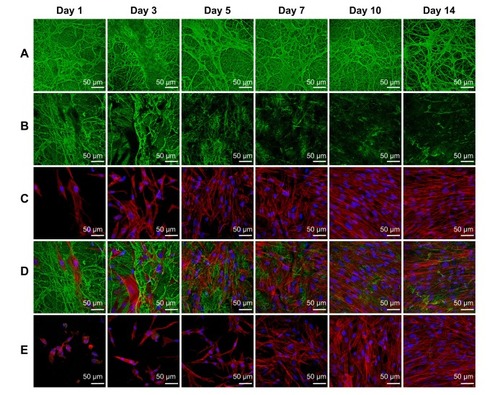
Figure 4 Mitochondrial activity in human dermal fibroblasts determined by an MTS assay in six time intervals (on days 1, 3, 5, 7, 10, and 14 after cell seeding) on nonmodified PLA membranes or on PLA membranes with a fibrin nanocoating (F).
Notes: Cells cultivated in the standard cell culture medium or in a medium supplemented with AA. The PSs were used as a control material. Arithmetic mean ± SD from 16 measurements made on four independent samples for each experimental group and time interval. ANOVA, Student–Newman–Keuls method, statistical significance (P≤0.05): *compared with a nonmodified PLA membrane in the standard cell culture medium or in the medium with AA, and AA compared with membranes in the standard cell culture medium.
Abbreviations: PLA, polylactide; AA, 2-phospho-l-ascorbic acid trisodium salt; PSs, polystyrene culture dishes; SD, standard deviation; ANOVA, analysis of variance; PLA F, fibrin nanocoating on PLA.
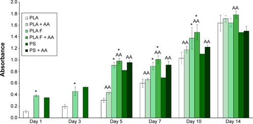
Figure 5 Morphology of human dermal fibroblasts on days 1 and 3 after seeding on nonmodified PLA membranes, or on PLA membranes with a fibrin nanocoating (F).
Notes: Cells cultivated in the standard cell culture medium. The PS was used as a control material. Cells stained with Texas Red C2-Maleimide and Hoechst #33258. Olympus IX 51 microscope, obj 10×, DP 70 digital camera.
Abbreviations: PLA, polylactide; PS, polystyrene culture dish; obj, objective.
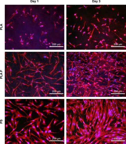
Figure 6 Morphology of human dermal fibroblasts on days 7 and 14 after seeding on nonmodified PLA membranes or on PLA membranes with a fibrin nanocoating (F).
Notes: Cells cultivated in the standard cell culture medium or in the medium supplemented with AA. The PS was used as a control material. Cells stained with Texas Red C2-Maleimide and Hoechst #33258. Olympus IX 51 microscope, obj 10×, DP 70 digital camera.
Abbreviations: PLA, polylactide; AA, 2-phospho-l-ascorbic acid trisodium salt; PS, polystyrene culture dish; obj, objective.
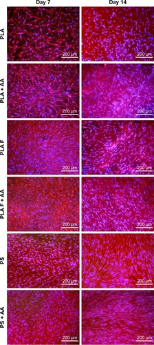
Figure 7 Relative expression of β1-integrins in human dermal fibroblasts on days 7, 10, and 14 after seeding on nonmodified PLA membranes or on PLA membranes with a fibrin nanocoating (F) determined by real-time PCR.
Notes: The cells were cultivated in the standard cell culture medium or in the medium supplemented with AA. The PS served as a control material. Reference gene GAPDH. ANOVA, Student–Newman–Keuls method, statistical significance (P≤0.05): *in comparison with the nonmodified PLA membrane in the standard cell culture medium or in the medium with AA.
Abbreviations: PLA, polylactide; PCR, polymerase chain reaction; AA, 2-phospho-l-ascorbic acid trisodium salt; PS, polystyrene culture dish; GAPDH, glyceraldehyde 3-phosphate dehydrogenase; ANOVA, analysis of variance.
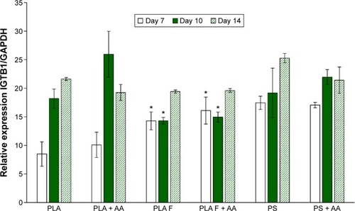
Figure 8 Immunofluorescence staining of β1-integrins in human dermal fibroblasts on day 3 after seeding on nonmodified PLA membranes or on PLA membranes with a fibrin nanocoating (F).
Notes: Arrows show focal adhesions containing β1-integrins. The cells were cultivated in the standard cell culture medium or in the medium supplemented with AA. Microscopic glass coverslips (GLASS) served as a control material. Cell nucleus stained with Hoechst #33258 (blue). Leica TCS SPE DM2500 confocal microscope, obj 63×/1.3 NA oil.
Abbreviations: PLA, polylactide; AA, 2-phospho-l-ascorbic acid trisodium salt; obj, objective; NA, numerical aperture.
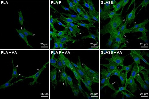
Figure 9 Relative expression of collagen I in human dermal fibroblasts on days 7, 10, and 14 after cell seeding on nonmodified PLA membranes or on PLA membranes with a fibrin nanocoating (F) determined by real-time PCR.
Notes: The cells were cultivated in the standard cell culture medium or in the medium supplemented with AA. The PS served as a control material. Reference gene GAPDH. ANOVA, Student–Newman–Keuls method, statistical significance (P≤0.05): *in comparison with the nonmodified PLA membrane in the standard cell culture medium or in the medium with AA, AA in comparison with membranes in the standard cell culture medium, and #statistical significance between PLA + AA and PLA F on day 14.
Abbreviations: PLA, polylactide; PCR, polymerase chain reaction; AA, 2-phospho-l-ascorbic acid trisodium salt; PS, polystyrene culture dish; GAPDH, glyceraldehyde 3-phosphate dehydrogenase; ANOVA, analysis of variance.
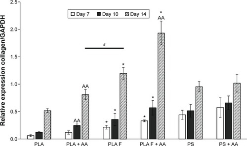
Figure 10 Immunofluorescence staining of extracellular collagen I fibers (green) produced by human dermal fibroblasts on days 7, 10, and 14 after seeding on nonmodified PLA membranes or on PLA membranes with a fibrin nanocoating (F).
Notes: The cells were cultivated in the standard cell culture medium or in the medium supplemented with AA. Microscopic glass coverslips (GLASS) served as a control material. Cell nucleus stained with Hoechst #33258 (blue). Leica TCS SPE DM2500 confocal microscope, obj 40×/1.15 NA oil.
Abbreviations: PLA, polylactide; AA, 2-phospho-l-ascorbic acid trisodium salt; obj, objective; NA, numerical aperture.
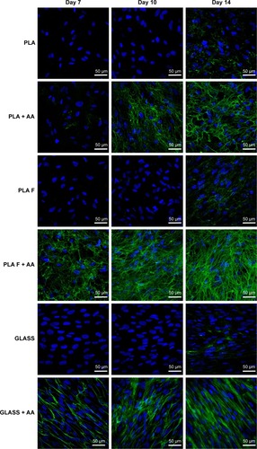
Figure 11 Immunofluorescence staining of total type I collagen (green) in human dermal fibroblasts on day 14 after cell seeding on nonmodified PLA membranes or on PLA membranes with a fibrin nanocoating (F).
Notes: The cells were cultivated in the standard cell culture medium. Microscopic glass coverslips (GLASS) served as a control material. Cell nucleus stained with Hoechst #33342 (blue). Leica TCS SPE DM2500 confocal microscope, obj 40×/1.15 NA oil.
Abbreviations: PLA, polylactide; obj, objective; NA, numerical aperture.
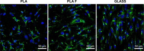
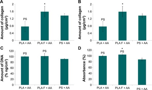
Figure 12 Total amount of type I collagen produced by human dermal fibroblasts on day 14 after seeding on nonmodified PLA membranes or on PLA membranes with a fibrin nanocoating (F).
Notes: Cells cultivated with AA added into the cell culture medium. The PS was used as a control material. Amount of collagen adjusted to the amount of cell DNA (A) or to the cell mitochondrial activity (B). Amount of cell DNA determined by a Picogreen assay (C). Cell mitochondrial activity determined by an MTS assay (D). Arithmetic mean ± SEM from 12 measurements made on four independent samples for each experimental group. ANOVA, Student–Newman–Keuls method, statistical significance (P≤0.05): *compared to a nonmodified PLA membrane, PS compared to the PS.
Abbreviations: PLA, polylactide; AA, 2-phospho-l-ascorbic acid trisodium salt; PS, polystyrene culture dish; SEM, standard error of mean; ANOVA, analysis of variance.