Figures & data
Figure 1 Typical computed tomography findings in idiopathic RPF. (A) Periaortic lesion (arrow) with aneurysmal change of the affected abdominal aorta and left hydronephrosis. (B) Right renal pelvic mass (arrow). (C) Right periureteral mass (arrow). (D) Placoid lesion (arrow) in the pelvis.
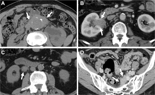
Figure 2 Proposed algorithm for the management of idiopathic RPF.
Abbreviations: GC, glucocorticoids; IS, immunosuppressants.
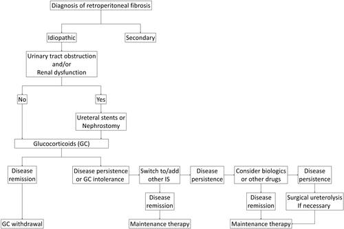
Table 1 Main Clinical Pictures, Laboratory Findings, Treatment, and Outcomes of Patients with IRPF in Different Clinical Series
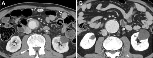
Figure 3 Aneurysm formation at the periaortic lesion of the abdominal aorta after glucocorticoid therapy. Before (A) and after (B) glucocorticoid therapy, the periaortic lesion is markedly ameliorated, while an aneurysm has developed at the same site.