Figures & data
Figure 1 Sensitivity and specificity of resistive index (RI) and delta RI in partial urinary obstruction.
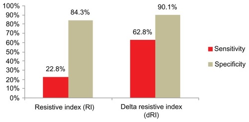
Table 1 Site of stone in the urinary tract of patients with unilateral ureteric obstruction
Table 2 Degree of pyelocaliceal dilatation on the side of urinary obstruction
Figure 2 Time period between symptom onset and Doppler ultrasound of the obstructed patients.
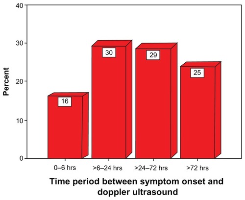
Table 3 Mean resistive index (RI) of obstructed and nonobstructed kidneys (comparison with several previous studies)
Figure 4 (A) Patient presents with acute right sided renal colic. Spectral waveform from the arcuate arteries of the upper pole of right kidney shows elevated RI of 0.714. Spectral waveform from the arcuate arteries of the mid pole of left kidney shows normal RI of 0.55. In this case both RI and delta RI are elevated.
Figure 4 (B) Subsequent IVU of same patient shows complete obstruction on right side due to a distal ureteral calculus.
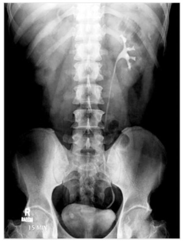
Figure 5 (A) Patient presents with acute left sided renal colic. Spectral waveform from the arcuate arteries of the both kidneys shows normal RI of 0.60 and 0.68. However delta RI is elevated.
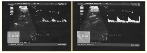
Figure 5 (B) IVU shows partial obstruction at left ureter due to a small calculus at left ureterovescial junction.
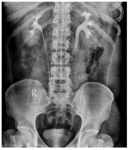
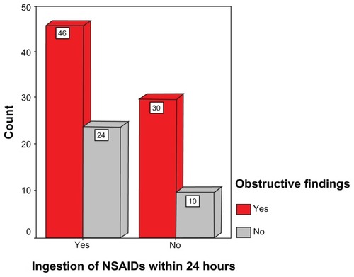
Figure 3 Effect of administration of nonsteroidal anti-inflammatory drugs (NSAIDs) on obstructive findings by resistive index.