Figures & data
Table 1 Classes of lupus nephritis
Table 2 Clinical indications for renal biopsy
Table 3 Distribution of cases within classes of lupus nephritis based on race
Table 4 Patient clinical information based on final diagnosis
Table 5 Case pathology findings for classes of lupus nephritis
Figure 1 Class I lupus nephritis. Mesangium and capillary loops appear normal in hematoxylin and eosin-stained section (A), Masson’s trichrome (B), periodic acid-Schiff (C) and methenamine silver (D). Transmission electron microscopy also shows no abnormality in the capillary loops (E) or mesangium (F).
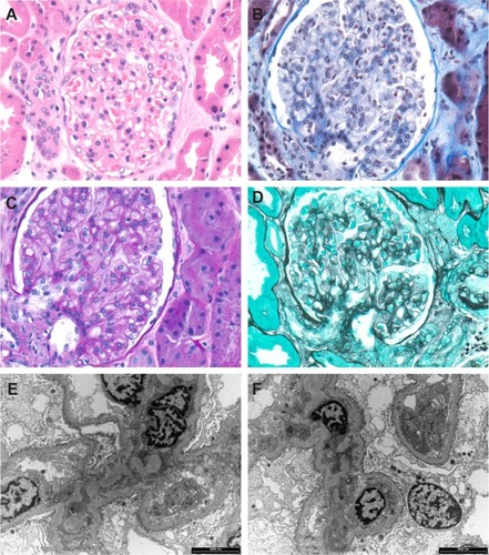
Figure 2 Glomerular capillary loop with granular trace deposits (arrows) in the subendothelium of the capillary basement membrane in a case with Class I lupus nephritis.
Abbreviations: EC, endothelial cell; P, podocyte.
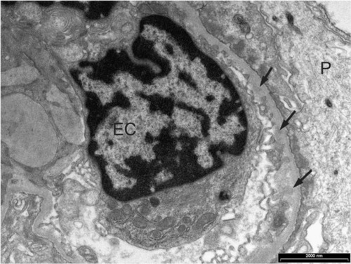
Figure 3 Class MB lupus nephritis.
Abbreviation: IgA, immunoglobulin A; IgG, immunoglobulin G; IgM, immunoglobulin M; KLC, kappa light chain; LLC, lambda light chain; C1q, complement 1q; C3c; complement 3c; Fib, fibrinogen.
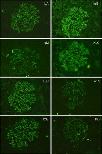
Figure 4 Class II lupus nephritis showing mesangial hypercellularity (asterisks).
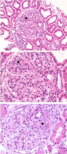
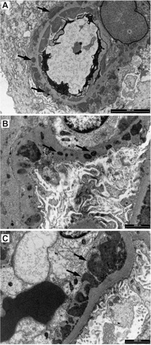
Figure 5 Granular electron dense deposits in Class MB lupus nephritis. (A) in the mesangium (short arrows) and (B) in the capillary basement membrane loops (long arrows).
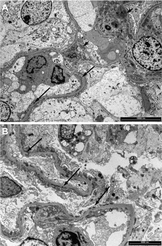
Figure 6 Class III lupus nephritis showing a case with typical spectrum and pattern of positive immunofluorescence staining.
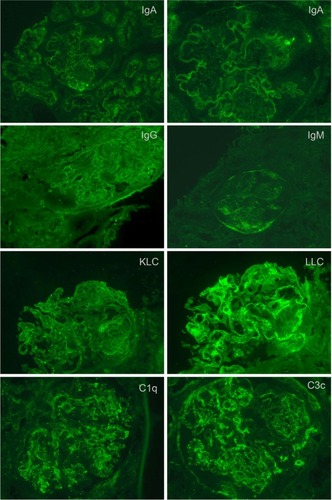
Figure 7 Electron microscopy showing (A) large fine granular deposits in the mesangial region (arrows), (B) prominent fine granular subendothelial deposits (arrows) and some intramembranous deposits (arrow heads) in a case of Class III lupus nephritis.
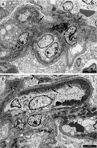
Figure 8 When tissue for immunofluorescence microscopy is not available, special histological stains and conventional immunohistochemical staining visualized with diaminobenzidine (DAB) can reveal glomerular deposits in Class III to Class VI. (A) Hematoxylin and eosin showing some crowding of nuclei in a segmental mesangial proliferative lesion; (B) Masson’s trichrome stain showing segmental mesangial proliferation and thickened capillary loops; (C) deposits of IgM (arrows); (D) deposits of IgG (arrows); (E) mesangial deposits of C3c (arrows); (F) deposits of C3c in capillary loops.
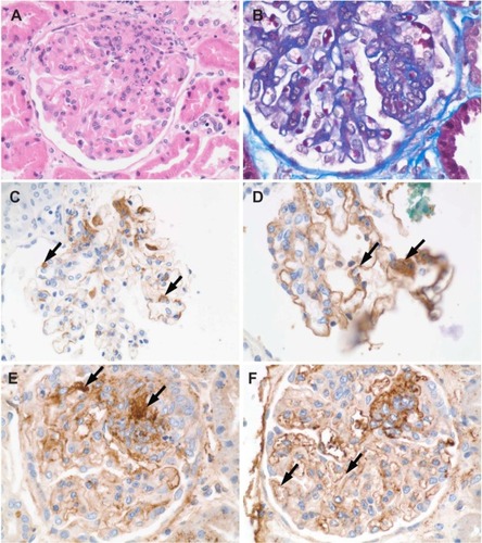
Figure 9 Class IV lupus nephritis. (A) Mesangial hypercellularity (asterisk) and deposits (arrows) associated with the capillary loops seen in a hematoxylin and eosin-stained section. (B) Foci of necrosis (asterisk), capillary loop deposits (arrow), and vascular thrombi (arrow head) are also seen. (C) Fuchsinophilic deposits (arrows) appear clearer in a Masson’s trichrome-stained section. (D) Hematoxylin bodies (arrows) appearing as darkly stained nuclei that blend into the background.
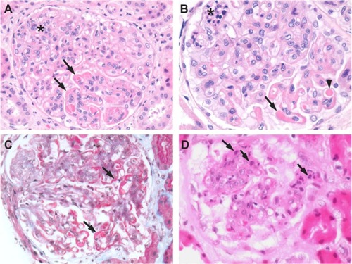
Figure 10 Class V lupus nephritis. Histological special stains are useful for further delineating the extent of glomerular deposits. (A) H&E-stained section of a Class V case showing mesangial hypercellularity. (B) H&E-stained section from a Class V case showing heavy deposits in the mesangium and capillary loops (arrows). (C) Fuchsinophilic deposits (arrows) on capillary loops seen in a Masson’s trichrome-stained section. (D) Deposits (arrows) in a Masson’s trichrome-stained section. (E) Deposits (arrows) seen in a PAS-stained section. (F) Basement membrane “spikes” in capillary loop deposits (arrows) seen in a methenamine silver-stained section.
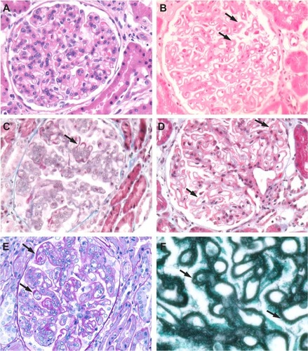
Figure 11 Class IV lupus nephritis.
Abbreviation: IgG, immunoglobulin G.
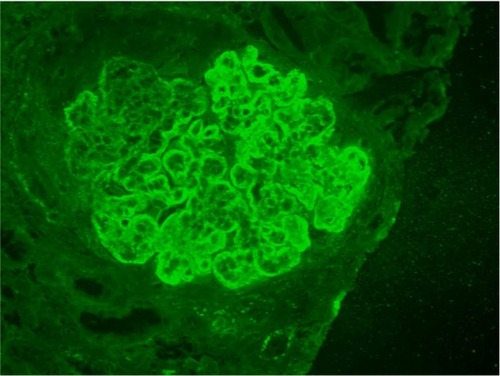
Figure 12 Class IV lupus nephritis large electron-dense granular deposits around capillary loops are seen in the subepithelial space (arrows in A), the intramembranous region of the basement membrane (arrows in B) and the subendothelial space (arrows in C).