Figures & data
Table 1 Initial Laboratory Results
Table 2 Full Immune Evaluation and Specific Tests
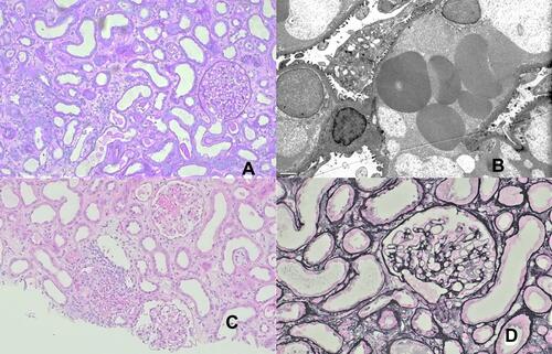
Figure 1 Kidney biopsy findings. (A) the glomerular compartment is unremarkable: normal capillary wall and no mesangial or endocapillary hypercellularity, renal cortex with inflammation ((A) periodic acid-Schiff stain, original magnification, 200x; (B) electron microscopy exhibiting podocyte focal foot process effacement without changes to the glomerular basement membrane; (C) renal cortex exhibiting mild interstitial edema and inflammation as well as acute tubular injury. (H&E stain, original magnification, 100x); (D) normal capillary walls without mesangial or endocapillary hypercellularity (Jones methenamine silver stain, original magnification, 200x).