
Figures & data
Figure 1 Original biopsy. The length of each bar represents the distance in microns. (A) shows dilated glomerular capillaries filled with mesh-like acellular material (Masson trichrome). (B) shows capillary lumina that are dilated with lipid droplets (Oil-Red-O stain). (C) shows a fingerprint-like structure composed of granules and vacuoles within a capillary lumen. There is expansion (asterisk) of the subendothelial space (electron microscopy, 5000x).
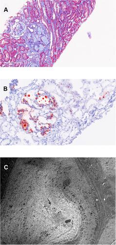
Figure 2 Follow up biopsy. The length of each bar represents the distance in microns. (A) shows glomerular capillaries are no longer dilated or filled with acellular material (Masson trichrome). (B) confirms the absence of lipid droplets (Oil-Red-O stain). (C) shows electron micrograph of capillary lumina that no longer contain acellular material (5000x). However, rare subendothelial spaces are expanded with lipoprotein material [osmiophilic granules (protein) and vacuoles (lipid)] as depicted in the inset (C) (15000x, corresponding to the rectangle on (C)).
![Figure 2 Follow up biopsy. The length of each bar represents the distance in microns. (A) shows glomerular capillaries are no longer dilated or filled with acellular material (Masson trichrome). (B) confirms the absence of lipid droplets (Oil-Red-O stain). (C) shows electron micrograph of capillary lumina that no longer contain acellular material (5000x). However, rare subendothelial spaces are expanded with lipoprotein material [osmiophilic granules (protein) and vacuoles (lipid)] as depicted in the inset (C) (15000x, corresponding to the rectangle on (C)).](/cms/asset/9073735a-3021-44b1-931a-dd5298a364a3/dnrd_a_12173917_f0002_c.jpg)