Figures & data
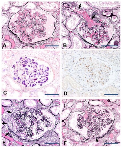
Figure 1 Renal pathology of DIC in association with metastatic prostate cancer. The peripheral glomerular capillaries (A) and afferent arteriole (B, arrows) were distended by thrombi formation (periodic acid methenamine silver stain). Fibrin strands revealed by phosphotungstic acid hematoxylin stain constituting the thrombi (C). The presence of sparsely scattered platelets within the thrombi was detected by immunostaining with an anti-CD41 antibody (D). Focal segmental sclerosis of the glomeruli (E and F; periodic acid methenamine silver stain). Segmental sclerosis of capillary architecture associated with attachments to the Bowman’s capsule with prominence of overlying epithelial cells (E, arrows). Segmental sclerosis was also noted at the urinary pole with capillary lumina obliterated by infiltration of mononuclear leukocytes and foam cells (F, arrowhead).