Figures & data
Figure 1 The flowchart of the study selection process.
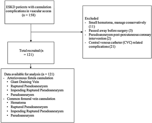
Figure 2 Clinical pictures of the cannulation complications from vascular access in ESKD patients: (A) Giant draining vein shown as a long pulsatile mass in the draining vein; (B) Ruptured pseudoaneurysm covered with a blood crust; (C) Impending rupture pseudoaneurysm with tense and glistening skin; (D) Pseudoaneurysm shown as a pulsatile mass without pain or bleeding; (E) Hematoma with bruise.
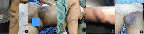
Table 1 Marfen and Hapsari Criteria for Cannulation Complications from Vascular Access in End-Stage Kidney Disease (ESKD) Patients
Figure 3 Ultrasound of the cannulation complications from vascular access in ESKD patients: (A) Giant draining vein shown as an enlarged draining vein more than 2 cm in diameter; (B) Acute hematoma visible as hypoechoic appearance without internal blood flow during color Doppler; (C) The yin-yang sign on Doppler ultrasound within the pseudoaneurysm.
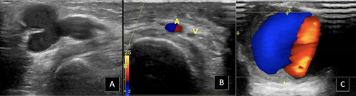
Figure 4 The Marfen and Hapsari flowchart of the surgical procedure for cannulation complications from vascular access in ESKD patients.
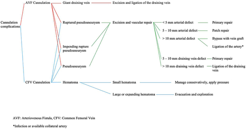
Table 2 Characteristics of Research Subjects
Table 3 ANOVA Analysis Between Diagnosis and Mean Differences of Subjective Symptoms (Days)
Table 4 ANOVA Analysis Between Diagnosis and Mean Differences of Hemoglobin and Leucocytes
Table 5 ANOVA Analysis Between Surgical Procedure and Mean Differences of Vascular Mass Diameter, Vascular Defect, and Hematoma Volume
Figure 5 Excised pseudoaneurysm and histological pictures of pseudoaneurysm and aneurysm: (A) An excised draining vein pseudoaneurysm; (B) Pseudoaneurysm histological examination showed a faint vascular appearance with numerous necrotic cells, hemosiderin deposits, macrophages, and stromal fibro-collagen tissue partially hyalinized with massive scattering of inflammatory cells, including lymphocytes, polymorphonuclear leukocytes, and eosinophils; (C) Aneurysm histological examination showed the tunica intima with a completely erosive endothelium and a fibro-collagenous fibrous connective tissue stroma covered with inflammatory cells, lymphocytes, and histiocytes; the tunica media is composed of partially fibrotic muscle cells with a normal nucleus; and the tunica serosa consists of connective tissue accompanied by mature adipose tissue with a normal nucleus.
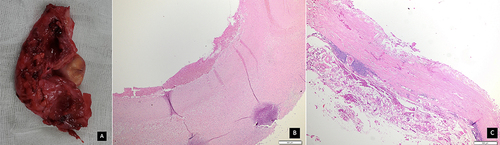
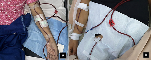
Figure 6 Cannulation of the AVF and the CFV: (A) The cannulation of the AVF, the outflow (red) placed on the proximal draining vein, and the inflow (blue) placed on the distal draining vein; (B) The cannulation of the CFV, the outflow (red) placed on the CFV, and the inflow (blue) placed on the cephalic vein.