
Figures & data
Figure 1 MRI of placenta increta (third pregnancy).
Notes: Coronal T2 MRI of the abdomen showing an irregular-looking posterior placenta, thinning of the myometrium, and discontinuity of the posterior uterine wall. A sigmoid loop (indicated by the arrow) is observed adjacent to the posterior uterine wall, however, it seems not to be invaded by the placenta. No dilation of the proximal or distal intestine was detected.
Abbreviation: MRI, magnetic resonance imaging.
Abbreviation: MRI, magnetic resonance imaging.
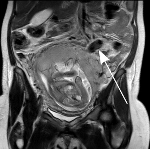
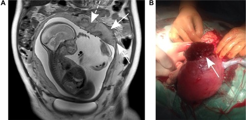
Figure 2 MRI of placenta percreta (fourth pregnancy).
Notes: (A) Coronal T2 MRI of the abdomen showing an irregular-looking posterior placenta, thinning of the myometrium, and discontinuity of the posterior uterine wall (arrows) with abnormal vascularity and prior hemorrhages (dashed arrow). (B) Placenta penetrating through 5×2 cm uterine wall rupture in the left cornu (arrow), as observed during the surgery.
Abbreviation: MRI, magnetic resonance imaging.
Abbreviation: MRI, magnetic resonance imaging.
Video S1 Sonography of placenta percreta (fourth pregnancy).
Notes: Transabdominal two-dimensional ultrasound in the transverse plane showing an abnormal placenta with thinning of the myometrium in the left posterior uterine wall, indicated by an asterisk. Similar findings are seen in the longitudinal plane in addition to fundal placental lacunas (arrow head) with increased blood flow by color Doppler scanning.