
Figures & data
Figure 1 (A) Color flow Doppler ultrasound showing velamentous umbilical cord inserting into fetal membranes, 2.19 cm off of placental disk for twin B in a monochorionic, diamniotic twin pregnancy identified at 20-week gestation. (B) Delivered monochorionic, diamniotic twin placenta at 30 weeks from same pregnancy as (A). There is a velamentous insertion of the umbilical cord for twin B (two clamps). Note the division of cord vessels within the fetal membranes and before they join the placental disk at multiple sites along the margin; insufficient imaging can confuse these vessels with a marginal cord insertion. The umbilical cord insertion site of twin A (one clamp) is inserted at the margin of the disk. This pregnancy was complicated by late-onset twin-to-twin transfusion syndrome with twin B, the donor, and twin A, the recipient.
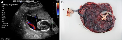
Figure 2 Color flow Doppler ultrasound from singleton pregnancy during a routine fetal anatomy survey showing the PCI to be centrally located on the placental disk.
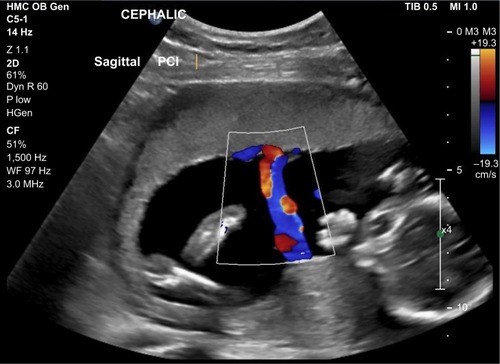
Figure 3 Flow diagram of study inclusion.
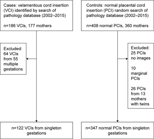
Table 1 VCI vs normal PCI, pregnancy comparisons
Table 2 VCI vs normal PCI, neonatal and placental comparisons
Table 3 Characteristics of umbilical cord identification at the time of fetal anatomy survey
Table 4 Performance characteristics of fetal anatomy survey in the diagnosis of VCI