Figures & data
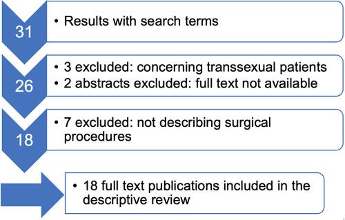
Figure 1 Intraoperative site during first surgery. The neovagina is attached to the promontory with PDS 2/0 sutures. After extensive dissection of adhesions there is no peritoneal coating present.
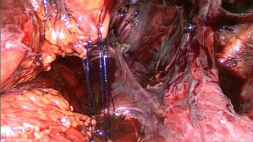
Figure 2 Initial intraoperative site during second surgery. The neovagina is hardly visible as it lays at the level of the pelvic floor and the former attachment to the promontory is detached completely.
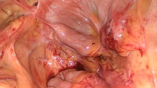
Figure 3 (A) During the second surgery a single mesh strip was placed over the apex of the neovagina. It was not possible to dissect the anterior compartment down to level of the bladder neck and at the posterior wall the vascular pedicle had to be spared. (B) The sketch illustrates schematically the course of the mesh and the relation of the vascular pedicle.
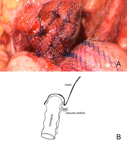
Figure 4 Vascular pedicle. Anteriorly the neovagina is visible. The strand deriving from the posterior wall corresponds to the vascular pedicle.
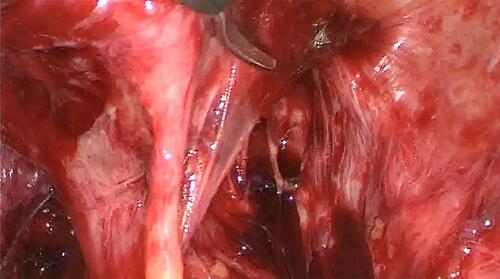
Table 1 Synopsis of surgical techniques for prolapse repair in the reported cases from the literature review
Figure 5 Procedure of literature selection with corresponding numbers of publications.