Figures & data
Figure 1 Normal amniotic fluid volume across gestation using quantile regression.
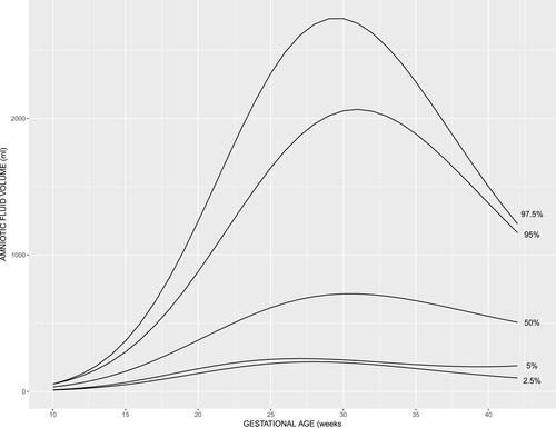
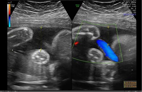
Figure 2 Example A of the assessment of amniotic fluid volume using gray scale on the L side of the screen and color Doppler on the R side of the screen and measurement of the amniotic fluid pocket.
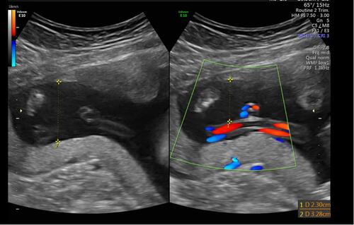
Figure 3 Example B of the assessment of amniotic fluid volume using gray scale on the L side of the screen and color Doppler on the R side of the screen and measurement of the amniotic fluid pocket.