Figures & data
Figure 1 The primary cultured cells were observed under an inverted microscope. Endometrial cells were completely adherent within 24 hours and oviduct mucosa cells adherent after 48 hours. Both cells were polygonal, tadpole and spindle type in primary culture and early passage. The nucleus was centered and round, and the cells gathered and grew like islands. Most of the cells began to change in morphology after passage to the third generation, and gradually appeared long fusiform. By the fifth generation, most of the cells grew into fusiform, similar to fibrous cells. (A) Primary endometrial cells; (B) primary oviduct mucosal cells; (C) the third generation of endometrial cells; (D) the third generation of oviduct mucosal cells; (E) the fifth generation of endometrial cells; (F) the fifth generation of oviduct mucosal cells.
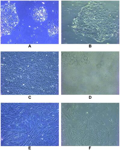
Figure 2 Immunofluorescence identification of epithelial marker keratin antibody was performed on endometrial cells and oviduct mucosa cells of the second generation. The results showed that 80–90% of the cytoplasm of endometrial cells and oviduct mucosa cells were stained with green fluorescence, showing positive cytokeratin antibodies. Microscopically, the cells were polygonal, spindle and spindle. Immunofluorescence identification of interstitial cell marker vimentin antibody was performed on endometrial cells and oviduct mucosa cells of the fifth generation. 80% of the cytoplasm of endometrial cells and oviduct mucosa cells were stained with red fluorescence under microscope. It showed positive cell vimentin antibody, and the cells were fusiform and polygonal under the microscope. (A1) The second generation of endometrial cells (DAPI). (A2) The second generation of endometrial cells (CK19). (A3) The second generation of endometrial cells (Merge). (B1) The second generation of oviduct mucosal cells (DAPI). (B2) The second generation of oviduct mucosal cells (CK19). (B3) The second generation of oviduct mucosal cells (Merge). (C1) The fifth generation of endometrial cells (DAPI). (C2) The fifth generation of endometrial cells (Vimentin). (C3) The fifth generation of endometrial cells (Merge). (D1) The fifth generation of oviduct mucosal cells (DAPI). (D2) The fifth generation of oviduct mucosal cells (Vimentin). (D3) The fifth generation of oviduct mucosal cells (Merge).
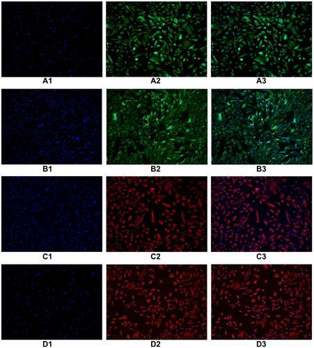
Figure 3 According to the same grouping method as mentioned above, the growth curve was drawn after 7 days of continuous measurement of endometrial cells and oviduct mucosa cells after co-culture. It can be seen that the growth rate of oviduct mucosa cells when growing alone was similar to that of endometrial cells in different proportions when co-culture, without significantly statistical difference (P > 0.05). The growth curve is of “S” type, which means that all of them experienced three stages: slow growth stage, logarithmic growth stage and plateau stage. TS: oviduct mucosal cells were cultured alone; T:E(1:1): the proportion of oviduct mucosal cells and endometrial cells was 1:1; T:E(1:0.5): the proportion of oviduct mucosal cells and endometrial cells was 1:0.5; T:E(1:0.1): the proportion of oviduct mucosal cells and endometrial cells was 1:0.1.
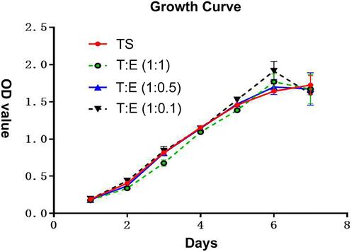
Figure 4 According to the same grouping method as mentioned above, endometrial cells were cocultured with oviduct mucosa cells and treated with decidualization. After 48 hours of culture, oviduct mucosa cells induced by decidualization began to show decidual-like changes, with large and round cell volumes, rich and transparent cytoplasm, and blurred cell boundaries. (A1) TS(40×), (A2) TS(100×), (A3) TS(200×), (B1) T: E(1:1)(40×), (B2) T:E(1:1)(100×), (B3) T;E(1:1)(200×), (C1) T:E(1:0.5)(40×), (C2) T:E(1:0.5)(100×), (C3) T:E(1:0.5)(200×), (D1) T:E(1:0.1)(40×), (D2) T:E(1:0.1)(100×), (D3) T:E(1:0.1)(200×).
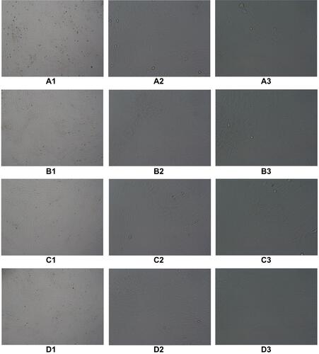
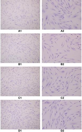
Figure 5 The cells in each group were treated with decidualization by HE staining, and the cytoplasm was stained eosin and the nucleus was blue. When oviduct mucosa cells were cultured alone and co-cultured with endometrial cells of different proportions, the cell morphology was basically unchanged, and all of them were fusiform, spindle and polygonal, a round, large and centered cell nucleus. (A1) TS(100×), (A2) TS(200×), (B1) T:E(1:1)(100×), (B2) T:E(1:1)(200×), (C1) T:E(1:0.5)(100×), (C2) T:E(1:0.5)(200×), (D1) T:E(1:0.1)(100×), (D2) T:E(1:0.1)(200×).