Figures & data
Figure 1 This is an endorectal ultrasound revealing a deep endometriosis nodule involving the muscular layer of the rectum.
Note: Copyright © 2013. Reproduced with permission of Elsevier. Roman H, Vassilieff M, Tuech JJ, et al. Postoperative digestive function after radical versus conservative surgical philosophy for deep endometriosis infiltrating the rectum. Fertil Steril. 2013;99(6):1695–1704.Citation62
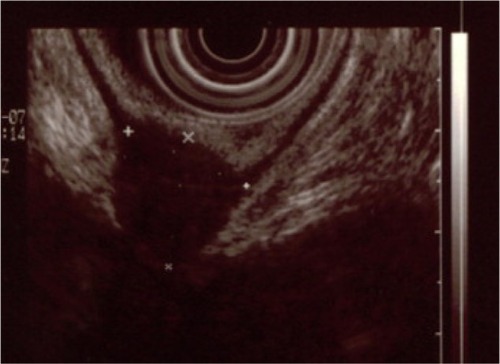
Figure 2 This is a modified virtual colonography with transparent views of the rectum and lower sigmoid.
Notes: The strictured area in the rectogenital area is indicated by arrows. The L, S, and D in the figure are from the LSD/MURO classification system. Copyright © 2013. Reproduced with permission of Elsevier. van der Wat J, Kaplan MD, Roman H, Da Costa C. The use of modified virtual colonoscopy to structure a descriptive imaging classification with implied severity for rectogenital and disseminated endometriosis. J Minim Invasive Gynecol. Epub June 5, 2013.Citation42
Abbreviations: L, length; S, stricture; D, distance to the anal verge; MURO, describes disseminated endometriosis beyond the rectogenital organs.
Abbreviations: L, length; S, stricture; D, distance to the anal verge; MURO, describes disseminated endometriosis beyond the rectogenital organs.
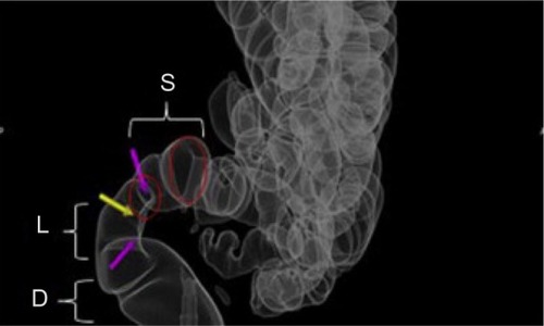
Figure 3 This is a preoperative assessment using magnetic resonance imaging, revealing a deep infiltrating endometriosis nodule with an obvious increase in rectal wall thickness.
Notes: The increase in rectal wall thickness is indicated by the arrow. Copyright © 2013. Reproduced with permission of Elsevier. Roman H, Vassilieff M, Tuech JJ, et al. Postoperative digestive function after radical versus conservative surgical philosophy for deep endometriosis infiltrating the rectum. Fertil Steril. 2013;99(6):1695–1704.Citation62
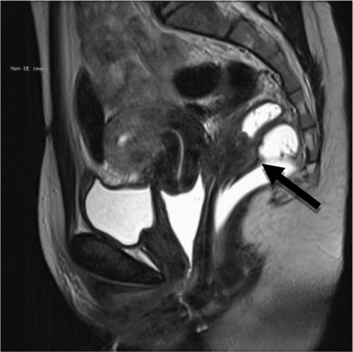
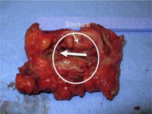
Figure 4 This is an endometriotic stricture >30% managed by segmental resection.
Note: Copyright © 2013. Reproduced with permission of Elsevier. van der Wat J, Kaplan MD, Roman H, Da Costa C. The use of modified virtual colonoscopy to structure a descriptive imaging classification with implied severity for rectogenital and disseminated endometriosis. J Minim Invasive Gynecol. Epub June 5, 2013.Citation42