Figures & data
Figure 1 Grayscale ultrasound image showing a cystic solid mass with clear boundaries in the posterior vaginal wall by abdominal ultrasonography (2020.05.23, (A)) and vaginal ultrasonography (2020.05.23, (B)).
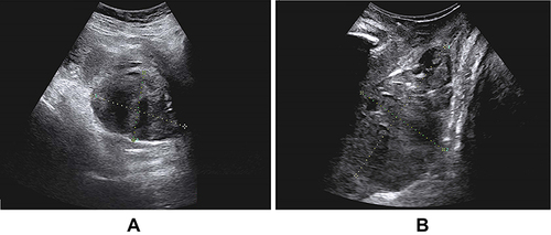
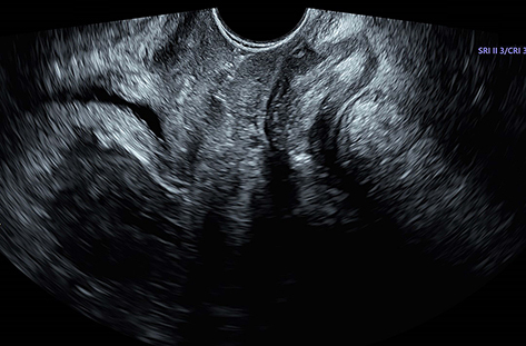
Figure 2 Grayscale ultrasound image showing a well-defined mass in the posterior vaginal wall (2020.05.25).
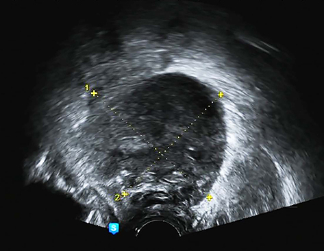
Figure 3 Vaginal ultrasound image showing a dark liquid area (2020.06.03).
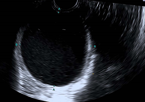
Figure 4 Vaginal ultrasound showing a dark, liquid area (2020.06.10).
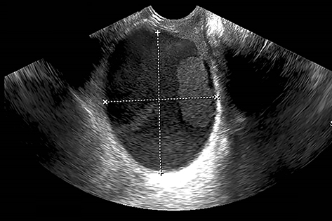
Figure 5 Grayscale ultrasound image showing the length of a mass (2020.06.15).
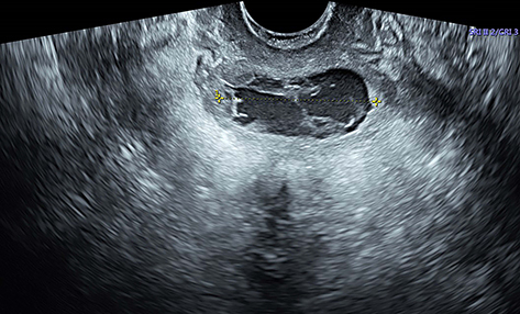
Figure 6 Normal vaginal ultrasound image indicating the disappearance of the mass (2020.07.03).