Figures & data
Figure 1 Bilateral mammography with nodular images in the left breast.
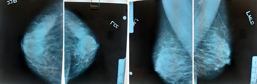
Figure 2 CT scan of thorax-abdomen-pelvis: (A) Hepatic tumor at the portal bifurcation; (B) Liver tumor at maximum diameter.
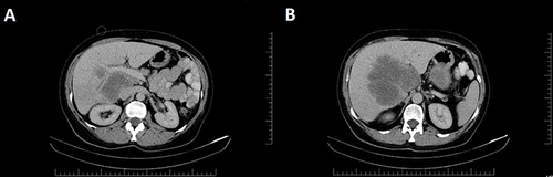
Figure 3 Bone scan: multiple bone metastases.
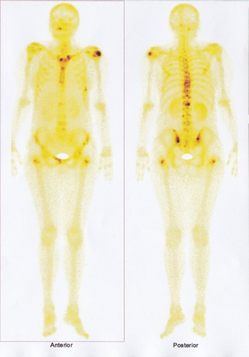
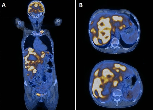
Figure 4 PET-CT: (A) overall image; (B) liver tumor.