Figures & data
Figure 1 The mean value in nulliparous and primiparous girls between 15 and 20 years old is ~40–50 cm3.
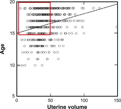
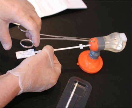
Figure 2 Illustration of how the fundal transverse diameter was measured with a specially designed instrument (cavimeter).
Figure 3 Adapted T-shape intrauterine device with transverse arm of 18 mm.
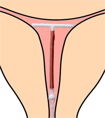
Table 1 Fundal transverse diameter (mm) according to age and parity (Kurz Cavimetric measurementsCitation8)
Figure 4 (A–C) Geometric relation of a properly inserted intrauterine device (IUD) to endometrial cavities with various inappropriate fundal transverse dimension. (A) Fundal transverse dimension significantly smaller than the length of the transverse arm of the IUD; (B) fundal transverse dimension significantly greater (initial position of the IUD); (C) fundal transverse dimension significantly greater (possible subsequent position of the IUD).
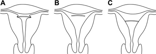
Figure 5 3-D coronal view of the uterine cavity, demonstrating the measurement of the fundal transverse dimension (19.0 mm).
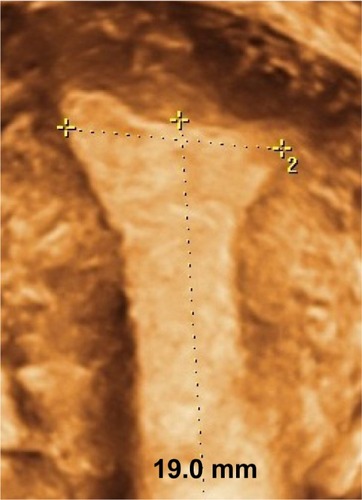
Table 2 Fundal transverse diameter (mm) according to gravidity/parity (3-D measurements)
Table 3 Number of women, gravidity/parity, and gross cumulative discontinuation rates per 100 users of GyneFix 330 and GyneFix 200 IUDs.
Figure 6 GyneFix 200 (Contrel Europe, Belgium), real size (left), in situ in foam uterus (middle), compared with the frameless FibroPlant-LNG (Contrel Europe, Belgium), which is derived from the frameless copper-releasing intrauterine device (IUD) (right).
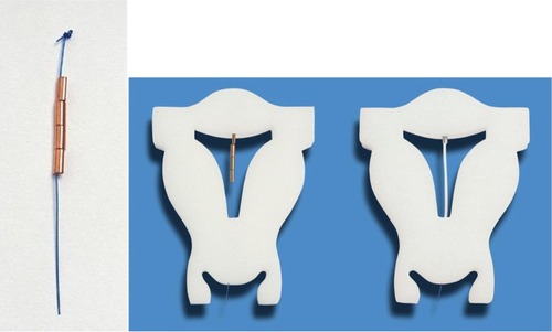
Figure 7 The current intrauterine device (IUD) is provided with a “visualized” anchor.
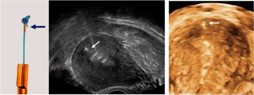
Figure 8 3-D ultrasonography: Abnormally located ParaGard intrauterine device (IUD) causing bleeding and pain (left) and middle (Mirena). Even if the IUD is apparently located in the correct position, the too-long transverse arm can cause painful contractions (right).
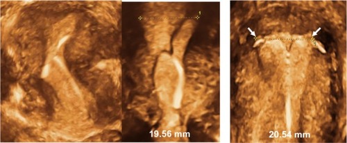
Figure 9 2-D/3-D ultrasonography: Position of the stem of the ParaGard intrauterine device (IUD), showing slight downward displacement (left). The arms of the IUD are unfolded and penetrate the muscular wall as the uterus is too small (right).
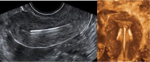
Figure 10 2-D/3-D ultrasonography: Another example of position of the stem of the Nova-T intrauterine device (IUD), showing slight downward displacement (left). The arms of the IUD are unfolded and penetrate the cornua of the uterus (middle, 3-D; right, hysteroscopy picture).
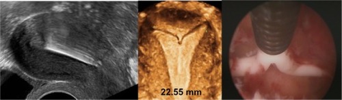
Figure 11 Mirena: shortened transverse arm after removal because of patient complaints and reinsertion under strict sterile conditions.
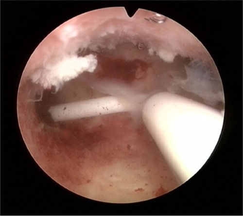
Figure 12 3-D ultrasound of GyneFix, illustrating the compatibility of the frameless intrauterine device with very narrow uterine cavities of young adolescent and nulliparous women.
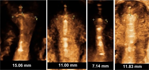
Figure 13 Home Uterine Trainer (HUT), suitable for home training of the frameless intrauterine device and intrauterine system insertion technique.