
Figures & data
Figure 1 Grossly, the uterine presented a spongy appearance with and necrosis and ulceration.
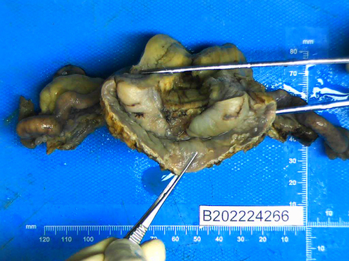
Figure 2 The tumor cells show vascular differentiation containing erythrocytes and mitosis (A)×40, (B)×100).
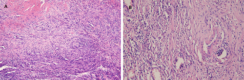
Figure 3 FISH showed YWHAE translocation proportion was 7 percent (≥30% is defined as positive).
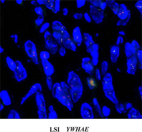
Table 1 Summary of Cases with Primary Angiosarcoma of the Uterine Cervix