Figures & data
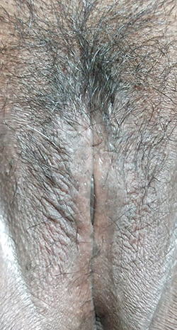
Figure 1 Clinical findings as pruritic multiple papules overlying the lichenified plaque prior to treatment.
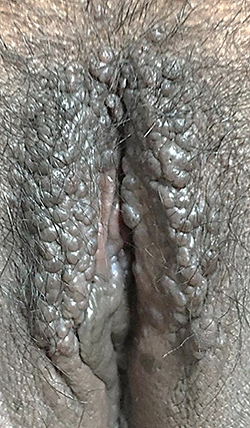
Figure 2 Histopathological examination from the labia majora revealed stromal lymphocytic infiltration.
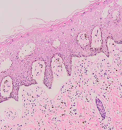
Figure 3 The result of immunohistochemical-staining. (A) Patchy p16 IHC staining was interpreted as p16-negative. (B) Weak and patchy p53 IHC staining was interpreted as p53-negative.
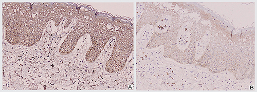
Figure 4 Clinical improvement was observed as the lesion became hyperpigmented macules without itching in the four weeks of follow-up.