Figures & data
Figure 1 The immunohistochemical staining results for p53 protein. (A) Wild-type p53, varying proportions and intensities of nuclear staining in tumor cells; (B) Aberrant nuclear overexpression of p53, strong nuclear staining in tumor cells compared to internal controls; (C) Complete loss of p53 expression, no p53 staining in tumor cell nuclei, with internal controls showing variable but moderate to strong staining. (D) Subclonal p53 mutation, with partial tumor cell cytoplasm showing diffuse moderate or strong positivity and no nuclear staining, accompanied by abnormal nuclear overexpression in some tumor cells.(Original magnification ×20).
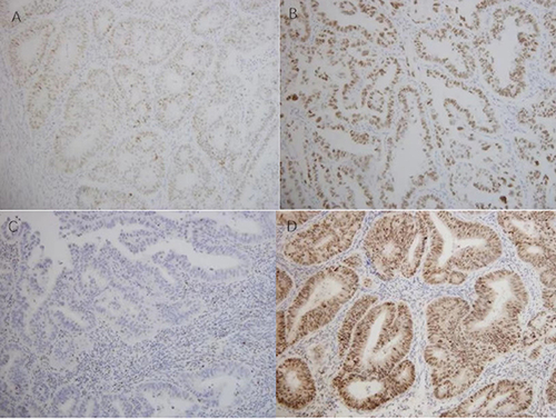
Figure 2 Recommended molecular typing detection pathway.
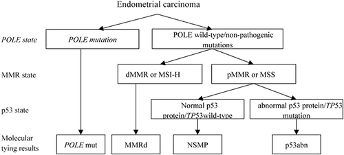
Table 1 Clinicopathological Characteristics of the Four Molecular Subtypes of Endometrial Carcinoma
Table 2 Correlation Analysis of Semi-Quantitative Parameters of Integrated PET/MRI in Early Endometrial Cancer Risk Stratification
Figure 3 The value of SUVmax in predicting the risk stratification of early endometrial cancer.
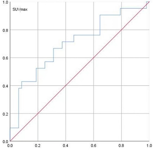
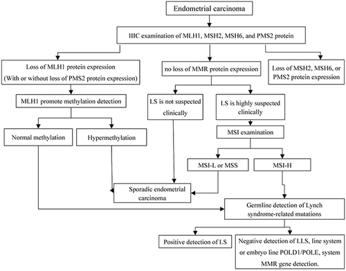
Figure 4 Lynch syndrome screening process.