Figures & data
Figure 1 Ultrasound findings of the first consultation in March 2022. (A) Cystic adenomyoma within the left uterine wall, filled with a content of ground-glass echogenicity. Hyperechoic, possibly calcified spots line the inside of the cyst wall. The measurement lines show a maximum diameter of 33.8 mm. (B) On color Doppler imaging no blood flow can be detected within the cystic adenomyoma, underlining its cystic nature. (C) The cystic adenomyoma is located in the left uterine wall and has no contact with the uterine cavity (green double arrows demonstrating the distance between the uterine cavity and the cystic adenomyoma). (D) The myometrium in the area of the lateral border of the cystic adenomyoma appears to be very thin (yellow arrows). (E) Image of the left ovary (red star) demonstrating that the cyst is not ovarian in origin in the sense of an endometrioma but located in the uterine wall. (F) Normally sited, intrauterine pregnancy to the patient’s right (Orange arrow). To the patient’s left, is the cystic adenomyoma.
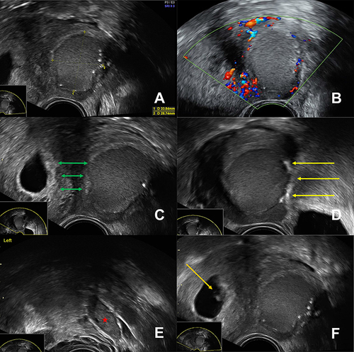
Figure 2 Transvaginal ultrasound in October 2022 shows the normally sited pregnancy within the uterine cavity to the patient’s right and, clearly separated from it, the cystic adenomyoma to the patient’s left. Its sonographic appearance is unchanged compared to the previous scans.
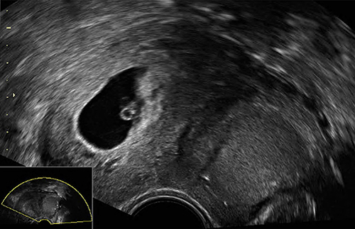
Figure 3 Transvaginal ultrasound in January 2023 during the second trimester of pregnancy, depicting the area of the anterior uterine wall to the left. In (A) and (B) the cystic adenomyoma is marked by the yellow measurement lines. It is practically only discernible by the marginally located hyperechoic spots (red arrows). The ground-glass content has dissolved. (C) and (D) show the peripheral blood vessels surrounding the cystic adenomyoma by color Doppler imaging. The hyperechoic spots indicate the former inner cyst wall (yellow arrows).
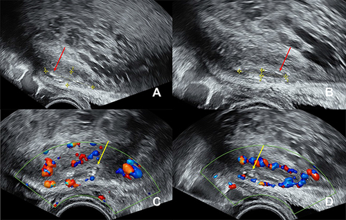
Figure 4 Transvaginal ultrasound in April 2023 during the third trimester of pregnancy, depicting the area of the anterior uterine wall to the left. The bladder, which is closest to the ultrasound probe, is almost empty. Cranial to the bladder is the part of the anterior uterine wall where the cystic adenomyoma was located on the previous scans (red arrow), however, at this point during pregnancy it is no longer visible. The caudal edge of the placenta and the fetal head are also visible.
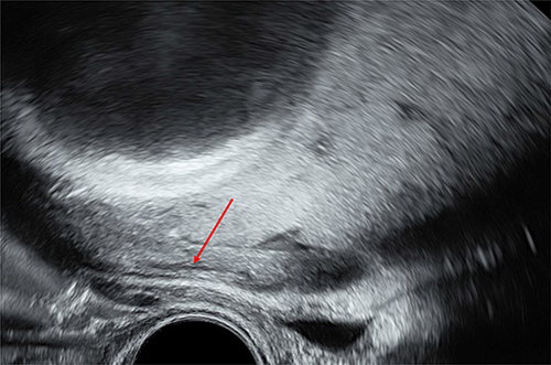
Figure 5 Intraoperative findings in May 2023. (A) and (B) show the intraoperative view of the anterior uterine wall at the level of hysterotomy during cesarean section. The cystic adenomyoma is not visible.
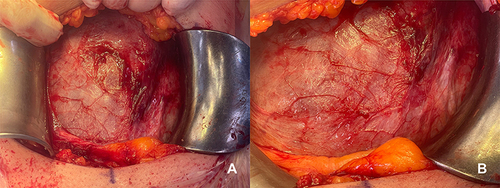
Figure 6 Transvaginal ultrasound in September 2023, four months postpartum. (A) Uterus depicted in the sagittal plane. The cystic adenomyoma appears as an unremarkable, ill-demarcated, hypoechoic area (red arrow) in the anterior uterine wall. (B) Located within the hypoechoic area are the characteristic white spots (yellow arrow). (C) Uterus depicted in the transverse plane. The cystic adenomyoma in the anterior uterine wall extends to the ventral surface of the uterus. (D) No vascularization can be detected within the hypoechoic area. The surrounding myometrium is well vascularized. The measurement lines indicate the dimensions of the cystic adenomyoma (10.8x6.7mm).
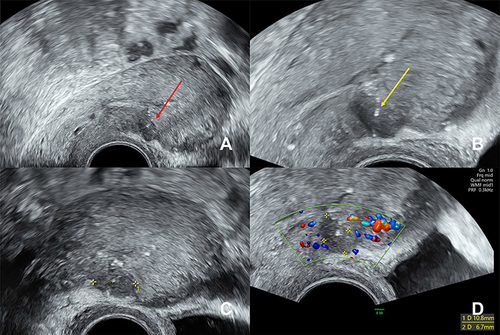
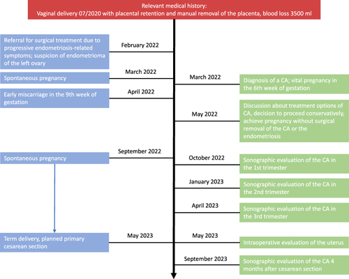
Figure 7 Diagram of the timeline for the episode of care.