Figures & data
Figure 1 (A) Combined transvaginal and transabdominal ultrasonogram showing a large irregular mass (13.1 cm × 8.2 cm) with hypoechoic features in the left side of the pelvic cavity; (B) Transvaginal ultrasonogram showing massive ascites in the abdominopelvic cavity.
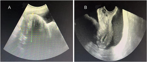
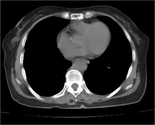
Figure 2 Chest computed tomography showing pleural fluid in both lungs.
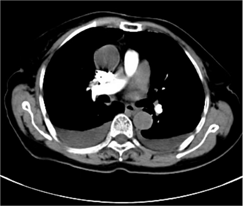
Figure 3 (A) Abdominal computed tomography revealed a soft tissue mass measuring approximately 10.9 × 8.2 × 14.3 cm with a clear boundary and heterogeneous density; (B) Contrast-enhanced scan showed no obvious enhancement and revealed that the supplying artery originated from the left ovarian artery.
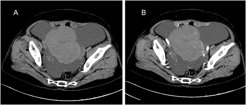
Figure 4 Histopathologic photomicrograph showing fibro cells and theca cells (hematoxylin and eosin staining, ×200).
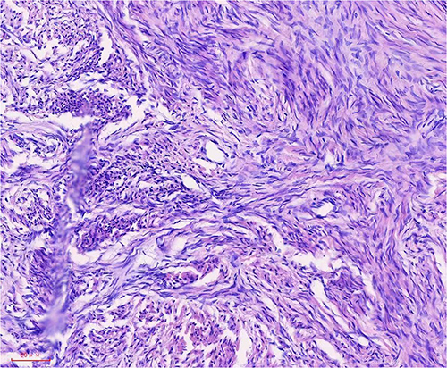
Figure 5 A follow-up chest computed tomography image 2 months after surgery showing resolution of the bilateral pleural effusion.