
Figures & data
Figure 1 Flow cytometry analysis of surface immune antigens on HUMSCs.
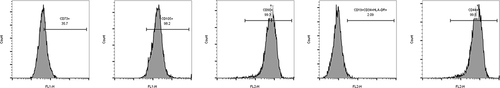
Figure 2 HUMSCs increases cell proliferation. (A) CCK-8 was used to detect cell proliferation; (B) The effect of HUMSCs on cell proliferation was analyzed by EDU assay, The nucleus is colored blue by DAPI and the cytoplasm is colored red by EDU.
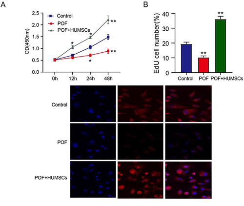
Figure 3 HUMSCs inhibit apoptosis of ovarian granulosa cells.
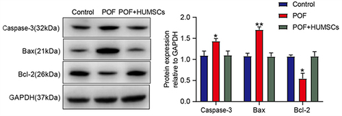
Figure 4 HUMSCs inhibit ovarian inflammation and oxidative stress induced by POF.
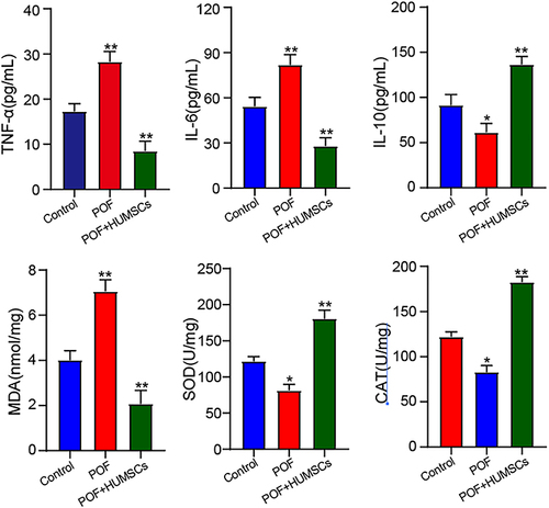
Figure 5 miR-10a promotes cell proliferation.
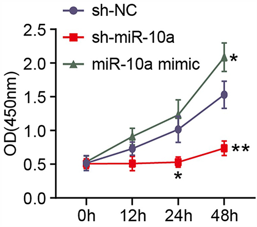
Figure 6 miR-10a-HUMSCs-Exosomes can be internalized into OGCs. (A) Red staining indicated PKH26-labeled miR-10a HUMSCs-exosomes. (B) Blue stains indicated DAPI-labeled nuclei. (C) Merged image of HUMSCs-exosomes uptake of miR-10a by GCs.
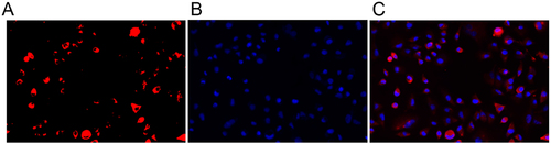
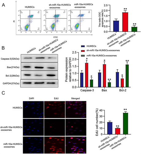
Figure 7 miR-10a-HUMSCs-exosomes increased the proliferation rate of POF OGCs and inhibited the apoptosis rate. (A) The impact of miR-10a-HUCMSCs-exosomes on OGC apoptosis was evaluated through flow cytometry analysis. (B) Protein expression was assessed through Western blot analysis; (C) The impact of miR-10a-HUCMSCs-exosomes on OGC proliferation was assessed using the EDU assay, The nucleus is colored blue by DAPI and the cytoplasm is colored red by EDU.
Data Sharing Statement
The simulation experiment data used to support the findings of this study are available from the corresponding author upon request.