Figures & data
Figure 1 The ultrasound imaging demonstrates a mass at the residual segment of the left fallopian tube (isthmus) consistent with an ectopic pregnancy.
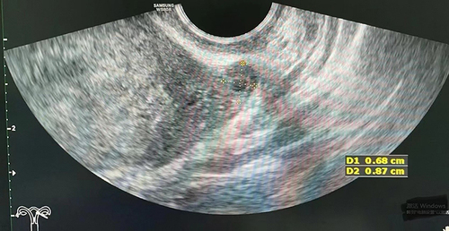
Figure 2 The size of the ectopic pregnancy mass in the left tubal remnant.
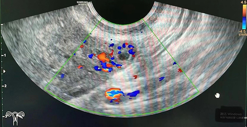
Figure 3 The blood flow pattern of the ectopic pregnancy mass.
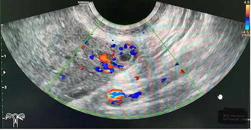
Figure 4 The pregnancy in the left tubal remnant was observed during laparoscopy.
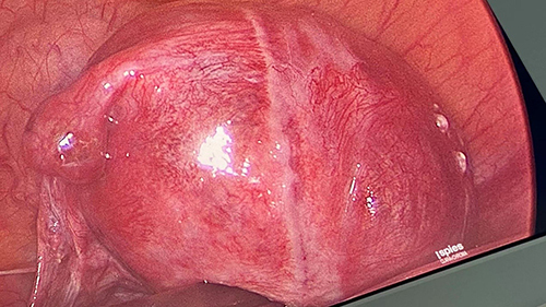
Figure 5 The right fallopian tube exhibits a normal appearance.
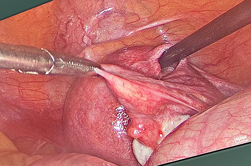
Figure 6 The villous tissue was present within the pregnancy sac of the left tubal remnant.
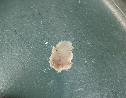
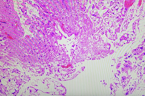
Figure 7 The pathological specimen confirmed chorionic villi (CV) within the ectopic pregnancy mass (haematoxylin and eosin stain, 200×magnification).