
Figures & data
Figure 1 Images of computerized tomography urography (CTU). There were no abnormalities in the shape and position of the right kidney, and no dilation of the right ureter. No normal kidney was found in the left renal area (A). An ectopically dysplasia kidney with peripheral enhancement could be seen anterior to fifth lumbar vertebra (B). Spotted contrast media was demonstrated around the ectopic kidney (thick arrow, (B). However, an enlarged uterus can be seen (C) and the thick-walled dysplasia kidney resembles an ovarian cyst (thick arrow, (D).
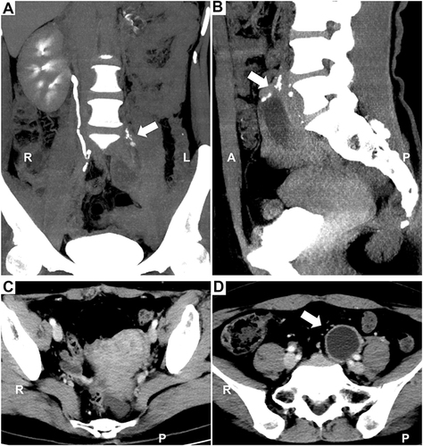
Figure 2 Images of MR. Sagittal turbo spin-echo (TSE) T2-weighted imaging (T2WI) (A-C), coronal T2WI (D-E), and axial T2WI with fat suppression (F). Sagittal and coronal T2WI showed an oval cyst in the right ovary (A and D). Another oval-shaped cystic lesion is observed above the uterus, with a long tubular tortuous downward extension on the left side of the pelvic cavity (B and E). However, careful examination revealed a maldeveloped ureter accompanying the uterus, the end of which was fused with the vagina (C and F). (Arrow: ectopic kidney; dotted line box: ectopic ureter).
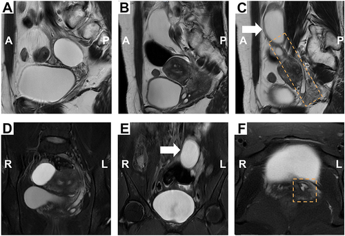
Figure 3 Intraoperative endoscopic data. (A) (a) Laparoscope showed septate uterus. A retroperitoneal cyst was dissociated from left ovary and fallopian tube. (b) The mass, probably be an ectopic kidney (arrow), was separated from surrounding tissue, and the distal end extended to nearby bladder. (B) Cystography imaging showed right ureter opening (dotted box) in bladder (a), but detected no left ureter opening (b).
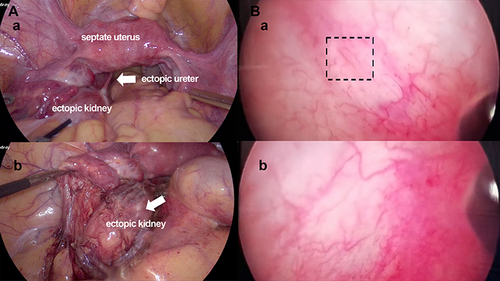
Figure 4 Gross of dysplasia ectopic kidney. Photograph shows the excised kidney with single system ureter.
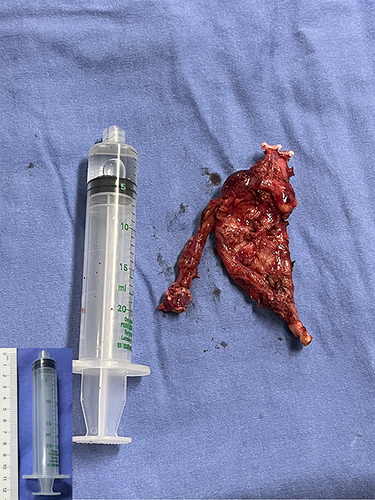
Table 1 Summary of Previous Publication of Ectopic Pelvic Kidney Mimicking Other Diseases
Table 2 Reports of Ectopic Kidney Combined with Ectopic Vaginal Ureter Causing Female Urinary Incontinence
Data Sharing Statement
Data used in the study are available from the corresponding author on request.