
Figures & data
Table 1 HLH-2004 Diagnostic Criteria and Patient’s Examination Results
Table 2 Patient’s Treatment Options and Temperature Changes
Figure 1 NK cell degranulation assay. PBMCs were isolated and co-incubated with a specific proportion of K562 cells for stimulation or incubated with medium alone (control group). Flow cytometry was then performed with anti-CD3 (FITC), anti-CD56 (APC) and anti-CD107a-PE labeled samples. CD3-CD56+NK cells were gated to assess their expression in response to K562 stimulation and non-stimulation by measuring amplitude changes in CD107a (NK-ΔCD107a) surface expression. The reference intervals were defined as <5% being deficient, >5% and <10% being abnormal, and >10% being normal.
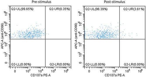
Figure 2 Bone marrow examination: active bone marrow hyperplasia, normal granulocyte lines with reactivity changes, elevated red blood cell lines, normal megakaryocyte lines, and observed tissue hemophagocytosis. (A) Bone marrow pathology (HE staining, ×80): bone marrow cells phagocytosis of nucleated red cells (as shown by arrow). (B) Bone marrow cytology (Reye’s staining, ×100 oil mirror): bone marrow cells phagocytosis of granulocytes and nucleated red cells.
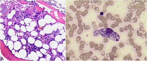
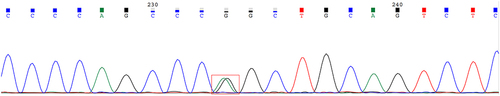
Figure 3 Exome sequencing revealed heterozygous mutations in UNC13D gene.
Data Sharing Statement
All supporting documents have been submitted along with the case report.