
Figures & data
Figure 1 Patient’s physical stature. (A) Patient has normal stature with height 170 cm; (B) Breast development shows Tanner 5; (C) Pubic hair development shows Tanner 4.
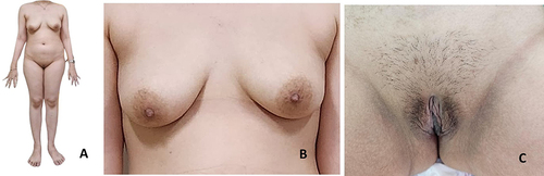
Figure 2 Patient’s abdominal ultrasonography. (A) Suspected solid ovarian mass with colour Doppler +1; (B) Free fluid present on Morrison pouch.
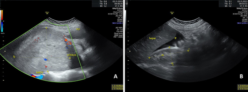
Figure 3 Patient’s chromosomal analysis. (A) Patient’s chromosome was 46XX; (B) FISH analysis reveals X mosaicism with no Y chromosome (X probe: red; Y probe: green).
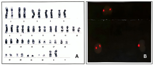
Figure 4 Intraoperative pictures. (A) Left ovarian mass; (B) Mass with uterus and right adnexa; (C) Ileum dilatation due to mass compression; (D) Mass after it was takien out, with estimated size of 20 x 15 x 15 cm, showing the uterus, right adnexa, peritoneal sampling, and omental sampling.
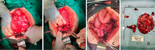
Table 1 Literature Review of 46XX Karyotype Patients with Germ Cell Malignancy