Figures & data
Figure 1 Selecting intracavitary versus interstitial brachytherapy.
Notes: (A) the red tumor at the time of diagnosis (left) reduces to a small size, and the green tandem and ovoids fit appropriately, so that the orange distribution of radiation dose adequately encompasses the residual disease. (B) a large red tumor at the time of diagnosis does not respond well, so an interstitial approach with multiple (green) little tubes is used to cover the full extent of residual disease, which would not have been possible using an intracavitary approach.
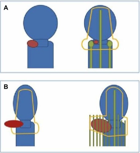
Figure 2 Examples of an intracavitary implant.
Notes: (A) the tandem, extending to the top of the uterine fundus. The ovoids are located at the level of the cervical os, and a white rectal paddle is noted posterior to the ovoids. The apparatus is held in place by a base plate. (B) three-dimensional rendering of the tandem and ovoid (top) and tandem and ring (bottom) in relation to the rectum and bladder. (C) coronal CT slice of the distribution of radiation using a tandem and ovoids (top) and tandem and ring (bottom).
Abbreviation: CT, computed tomography.
Abbreviation: CT, computed tomography.
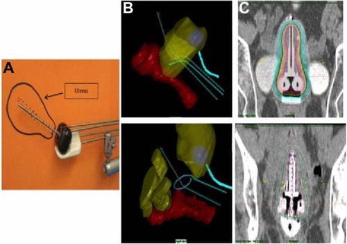
Figure 3 Examples of an interstitial implant.
Notes: (A) three-dimensional rendering of the organs at risk, in relation to the brachytherapy catheters and radiation dose cloud. (B) a completed implant. (C) axial CT slice showing the distribution of radiation dose achievable with an interstitial implant.
Abbreviation: CT, computed tomography.
Abbreviation: CT, computed tomography.
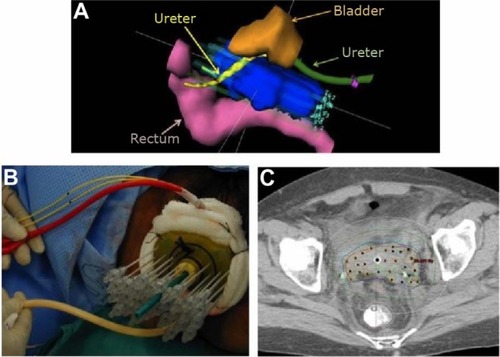
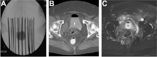
Figure 4 Examples of brachytherapy.
Notes: (A) X-ray. (B) axial CT slice. (C) axial MRI slice. One can see the increasing amount of anatomic detail that is apparent on MRI, compared with the other imaging modalities.
Abbreviations: CT, computed tomography; MRI, magnetic resonance imaging.
Abbreviations: CT, computed tomography; MRI, magnetic resonance imaging.