
Figures & data
Figure 1 Contrast-enhanced CT images performed on August 7 (A), August 14 (B), August 22 (C), and September 26 (D) showing change of isolated splenic vein thrombosis (red arrow).
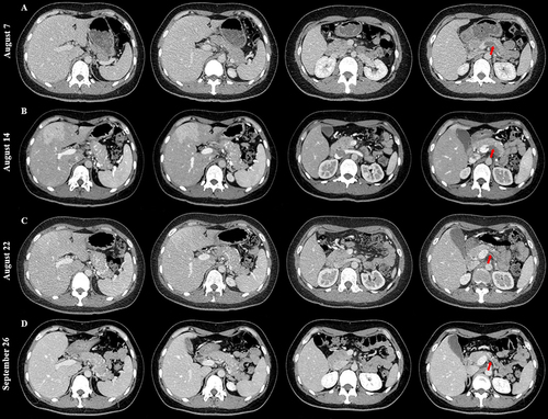
Figure 2 Upper gastrointestinal endoscopic images. There were no varices in the esophagus (A) or gastric fundus (B).
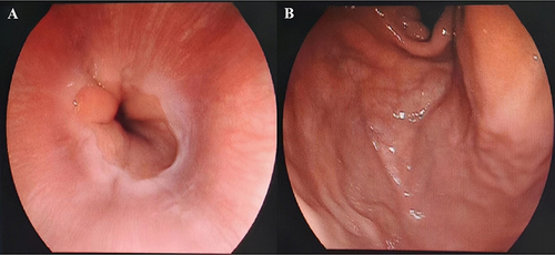
Table 1 Reported Cases of Isolated Splenic Vein Thrombosis or Occlusion: An Overview