Figures & data
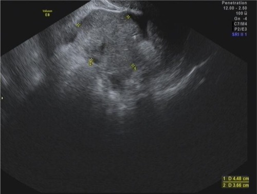
Figure 1 Transvaginal ultrasound in a 25-year-old woman.
Notes: This simple cyst measuring 64 mm by 42 mm was seen on transvaginal ultrasound in a 25-year-old woman complaining of lower abdominal pain. She was followed for several months with cyst persistence and therefore underwent laparoscopic cystectomy. On laparoscopy, a smooth walled cyst containing clear citrine fluid was seen. Pathology revealed a benign cystadenoma.
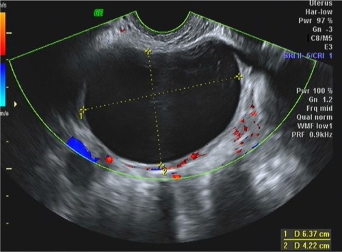
Figure 2 Transabdominal ultrasound in a 16-year-old adolescent.
Notes: A hemorrhagic cyst consistent with a corpus luteum cyst diagnosed on transabdominal ultrasound in a 16-year-old adolescent who presented with acute abdominal pain. A hypoechogenic cyst with an echogenic structure representing the blood clot is observed. In addition, the delicate “cobweb” is seen.
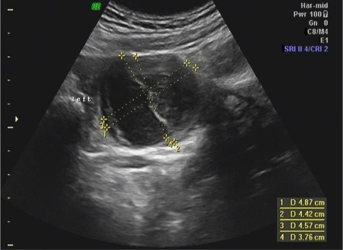
Figure 3 Transvaginal ultrasound in a 70-year-old woman.
Notes: A 90 mm dermoid cyst diagnosed on routine transvaginal ultrasound in a 70-year-old woman. The cyst contains mostly echoic material and produces a noticeable acoustic shadow with attenuation of the sound beyond the cyst.
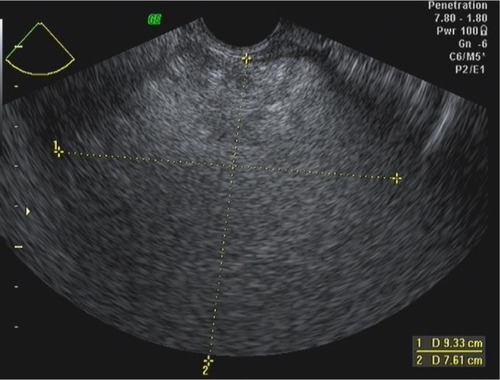
Figure 4 Transabdominal scan in a 9-year-old girl.
Notes: An enlarged ovary was seen on transabdominal scan in a 9-year-old girl who presented with abdominal pain. The ovary contained two cystic areas, one with an echoic structure. In addition, the stroma of the ovary appears edematous and the normal follicular structure is lost. On laparoscopy, torsion of the ovary involving a large dermoid cyst was diagnosed.
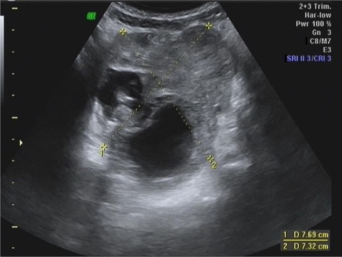
Figure 5 Transvaginal ultrasound in a 28-year-old nulligravida.
Notes: A tubular hypoechoic mass with indentations along it walls consistent with a hydrosalpinx was seen on transvaginal ultrasound in a 28-year-old nulligravida with known tubal occlusion on hysterosalpingogram. Laparoscopy confirmed these findings along with severe pelvic adhesions.
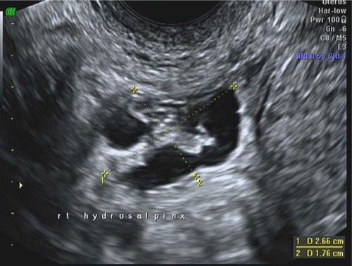
Figure 6 Ovarian cyst observed on transvaginal ultrasound in a 25-year-old woman.
Notes: A 58×44 mm ovarian cyst was observed on transvaginal ultrasound in a 25-year-old woman who presented with pelvic pain. The “ground glass” typical appearance of endometrioma is noticed. Laparoscopy confirmed the diagnosis.
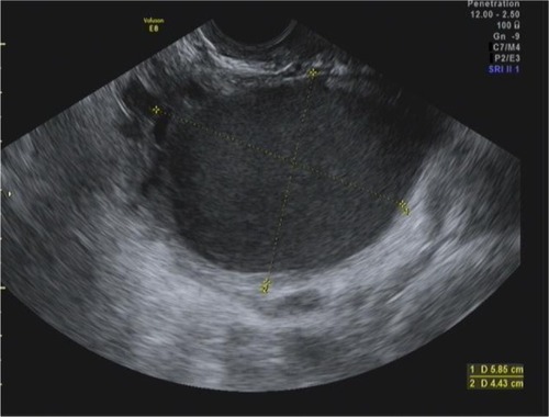
Figure 7 Transabdominal ultrasound scan in an 8-year-old girl with abdominal pain.
Notes: An enlarged ovary with loss of follicular structure was seen in an 8-year-old girl who presented with pelvic pain and vomiting. Laparoscopy confirmed torsion of the adnexa.
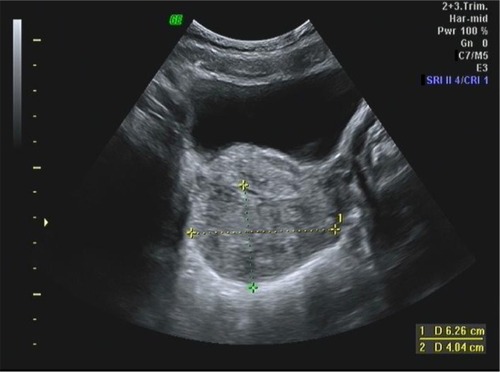
Figure 8 Transvaginal ultrasound in a 64-year-old woman with pelvic mass.
Notes: A pelvic mass measuring 83×95 mm and containing septations and papillations was seen in a 64-year-old woman. Surgery revealed an adenocarcinoma of the ovary.
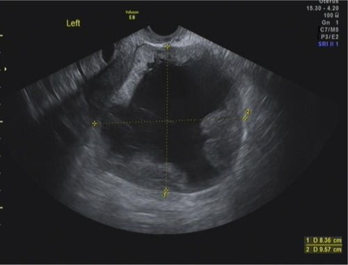
Figure 9 Transabdominal ultrasound scan in a 41-year-old woman.
Notes: This large cyst in a 41-year-old woman contains thin septations without increased Doppler blood flow. Surgery revealed a benign mucinous cystadenoma.
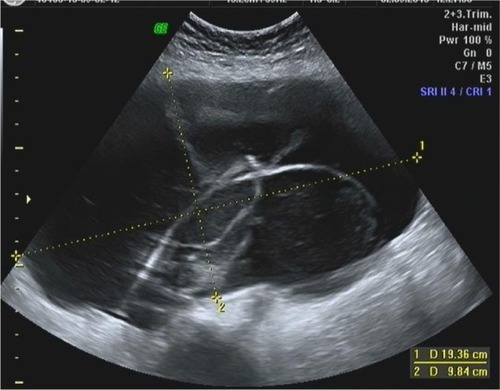
Figure 10 Transvaginal ultrasound scan of a 59-year-old woman.
Notes: The ovary of a 59-year-old woman contains a large solid mass. Surgery revealed this mass to be a benign fibrothecoma.