
Figures & data
Figure 1 Brain contrast-enhanced magnetic resonance imaging (MRI) scans show brain metastases before and after anlotinib and T-DXd therapy. (A) brain metastases before anlotinib and T-DXd therapy; (B) partial response in brain metastases after anlotinib and T-DXd treatment.
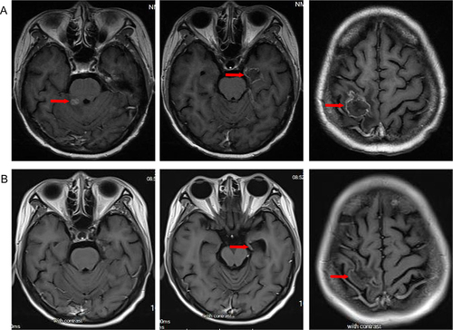
Figure 2 Chest and abdominal enhanced computed tomography(CT) scans show lung metastases before and after anlotinib and T-DXd therapy. (A) lung metastases before anlotinib and T-DXd therapy; (B) partial response in lung metastases after anlotinib and T-DXd treatment.
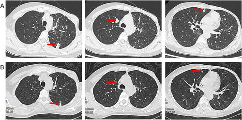
Figure 3 Timeline of the clinical course in this patient.
Abbreviations: NAT, Neoadjuvant therapy; AT, adjuvant therapy; M, months; ECMs, extracranial metastases; BMs, brain metastases; PR, partial response; PD, progress disease; SD, stable disease; THP, albumin-bound paclitaxel, trastuzumab and pertuzumab; WBRT, whole brain radiotherapy; HP, trastuzumab and pertuzumab; Cap, capecitabine; Bav, bevacizumab; NVB, navelbine; SBRT, stereotactic radiotherapy; RC48, disitamab vedotin; T-DXd, trastuzumab deruxtecan.
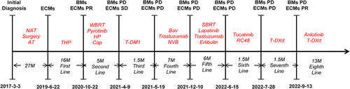
Table 1 Summary of Current Clinical Trials Using Targeted Agents in Her2+ BCBM