Figures & data
Figure 1 The four breast parenchymal patterns.
Notes: (A) Fatty; (B) scattered; (C) heterogeneously dense; (D) extremely dense.
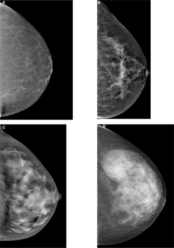
Figure 2 Diagram of the digital breast tomosynthesis (DBT) system.
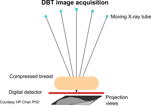
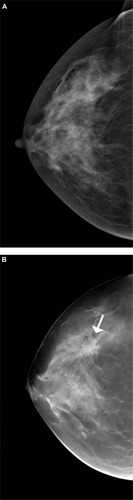
Figure 3 Cancer more evident with tomosynthesis.
Notes: (A) Craniocaudal view of screening digital mammogram. No abnormality is evident. (B) Digital breast tomosynthesis slice of the same patient, in the same position, depicts a spiculated mass in central breast not evident in the screening mammogram view. Proven invasive ductal carcinoma (arrow).