Figures & data
Table 1 The clinical presentation of uterine leiomyomas
Figure 1 A 37-year-old Afro-Caribbean woman with a history of menorrhagia. Transvaginal ultrasound image showed a bulky fibroid uterus with multiple intramural fibroids. The largest fibroid (pictured above) is at the fundus on the posterior wall measuring 4.5 × 4 cm.
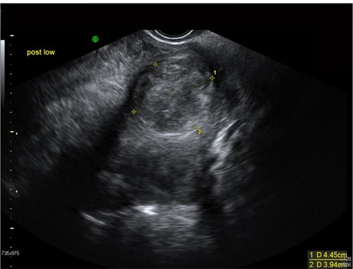
Figure 2 B-mode image (A) obtained during saline infusion sonohysterography in a 46-year-old woman with a diagnosis of an endometrial polyp and corresponding strain image (B) obtained using the 2-dimensional multilevel hybrid algorithm. Red arrows indicate the location of the polyp, and yellow contours indicate the outer uterine wall.
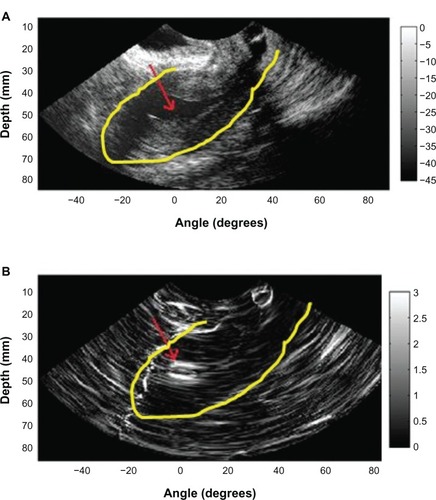
Figure 3 A 47-year-old woman with a hugely enlarged uterus. The fundus is at the level of L3. It contains a solitary large intramural fibroid which is mostly right sided, displacing the cavity to the left. This fibroid measures approximately 14.2 × 10.9 × 8.8 cm.
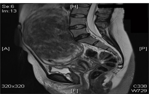
Figure 4 Mechanisms of action of progesterone receptor modulators on uterine fibroids.
Abbreviations: ER, estrogen receptors; PR, progesterone receptors; AR, androgen receptors; TRAIL, TNF-related apoptosis-inducing ligand; MMP, matrix metalloproteinases; TIMP, tissue inhibitors of metalloproteinases.
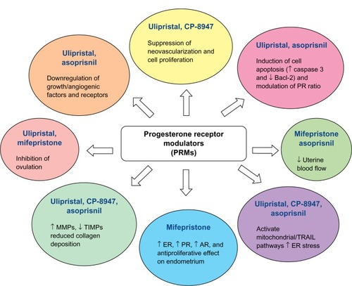
Figure 5 The UAE procedure. Drawings (A and B) illustrate the path of the catheter to deliver embolization particles to occlude the uterine arteries. Angiograms show the fibroid blood supply before (C) and after (D) UAE.
Abbreviation: UAE, uterine artery embolization.
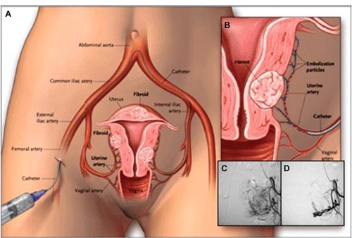
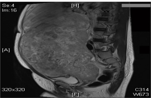
Figure 6 A 40-year-old woman with a large abdomino-pelvic mass, which turned out to be an aggressive leiomyosarcoma.