Figures & data
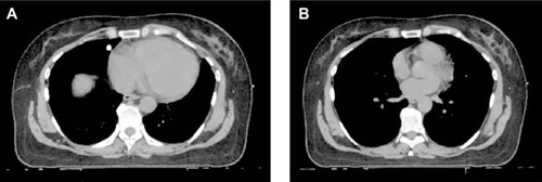
Figure 1 Deep inspiration breath hold (DIBH).
Notes: Representative axial computed tomography images of a patient undergoing treatment for left-sided breast cancer with (A) free breathing and (B) DIBH techniques. It is apparent that the lung volume has expanded and the heart has moved posteriorly, away from the chest wall.