Figures & data
Figure 1 Sagittal MRI of a typical case pre/post treatment.
Notes: (A) Sagittal T2-weighted image (T2WI) demonstrating a single uterine fibroid of predominantly low signal intensity, accessible by the ultrasound transducer, which is imbedded in the magentic resonance table. (B) Sagittal T1-weighted image (T1WI) postcontrast at screening demonstrates heterogeneous enhancement of the fibroid. (C) Sagittal T1WI postadministration of intravenous contrast after MRgFUS treatment demonstrates lack of enhancement in the fibroid, consistent with treatment effect.
Abbreviations: MRgFUS, magnetic resonance-guided focused ultrasound; MRI, magnetic resonance image.
Abbreviations: MRgFUS, magnetic resonance-guided focused ultrasound; MRI, magnetic resonance image.
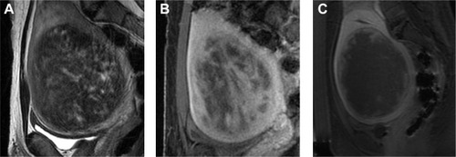
Figure 2 Examples of MRgFUS screen failures.
Notes: (A) Sagittal T1-weighted image (T1WI) prior to contrast demonstrates area of very low signal along the inferior margin of the fibroid. (B) After administration of contrast agent, there is lack of enhancement in the majority of the fibroid, and the clumpy areas of low signal are again seen. Both of these factors – lack of enhancement and clumpy low signal areas consistent with peripheral calcifications – are reasons for screen failure for an MRgFUS treatment. (C) Sagittal T1WI postadministration of intravenous contrast in a different case demonstrating complete lack of enhancement in two uterine fibroids; this case was also a screen failure.
Abbreviation: MRgFUS, magnetic resonance-guided focused ultrasound.
Abbreviation: MRgFUS, magnetic resonance-guided focused ultrasound.
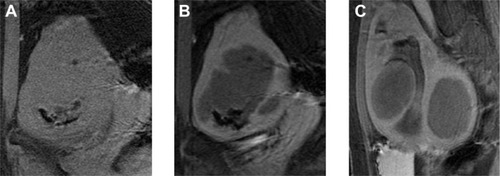
Figure 3 Example of MRgFUS screen failure due to innumerable small fibroids.
Notes: (A) Axial, (B) coronal, and (C) sagittal T2-weighted image of a patient with innumerable, small uterine fibroids, without a definite dominant fibroid. Cases such as these are not good candidates for MRgFUS.
Abbreviation: MRgFUS, magnetic resonance-guided focused ultrasound.
Abbreviation: MRgFUS, magnetic resonance-guided focused ultrasound.
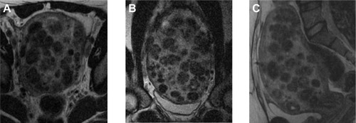
Figure 4 Illustrative example of cellular fibroid.
Notes: (A) Axial, (B) coronal, and (C) sagittal T2-weighted image (T2WI) demonstrating a fibroid of high signal intensity on T2WI. These fibroids are thought to represent very cellular fibroids, and may be difficult to treat.
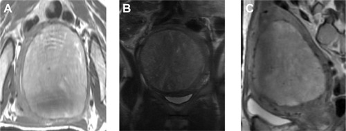
Table 1 MR imaging findings that require evaluation on the screening MR and on the planning MR on treatment day
Figure 5 Photograph depicting a patient lying in the prone position on the magnetic resonance (MR) table.
Notes: The focused ultrasound transducer array is located within the MR table, and the patient is positioned such that the fibroid overlies the ultrasound transducer. Reprinted with permission from InSightec Inc.

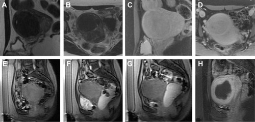
Figure 6 Illustrative example of manipulation of beam path obstruction.
Notes: (A) Screening sagittal T2-weighted image (T2WI) and (B) axial T2WI of an accessible solitary uterine fibroid, which demonstrated enhancement postadministration of intravenous contrast agent on sagittal and axial T1 postcontrast images (C, D). However, on the day of treatment, sagittal localizing images demonstrated multiple bowel loops anterior to the uterus (E). The bowel loops were manipulated by filling the bladder with saline and filling the rectum with ultrasound gel (F, sagittal localizer), which displaced the bowel loops superiorly to create a sonication window. After the bladder was emptied, the sonication window remained (G, sagittal localizer), allowing for satisfactory treatment (H, sagittal T1-weighted image (T1WI) postadministration of contrast agent) with a large area of nonenhancement.