Figures & data
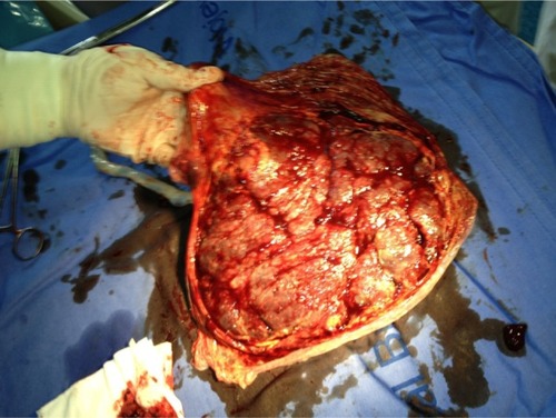
Figure 1 A gray-scale ultrasound image shows a well-defined circumscribed ovoid heteroechogenic placental mass with a 4.99 cm diameter.
Note: The mass protrudes from the fetal surface of the placenta and is in contact with the amniotic cavity.
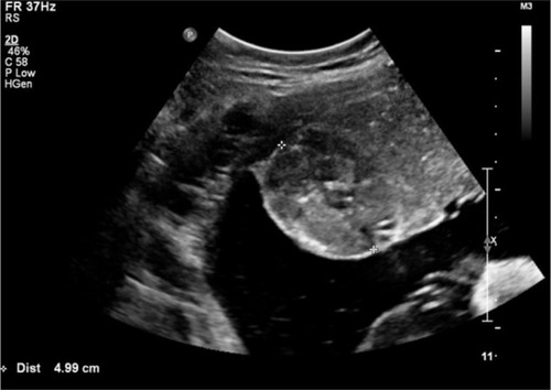
Figure 2 A gray-scale ultrasound image shows the protruding placental mass into the amniotic cavity from a placenta with the thickest anteroposterior diameter of 5.5 cm.
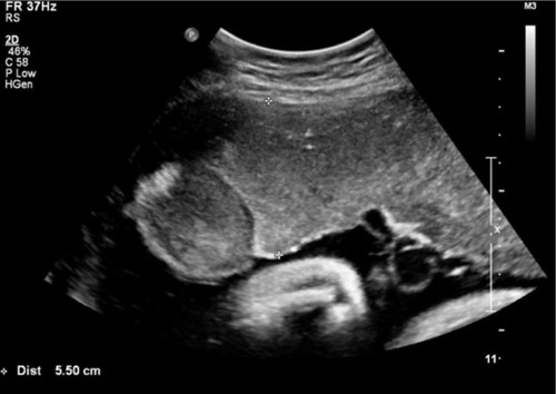
Figure 3 Macroscopic appearance of the placenta after normal vaginal delivery.
Note: A 5-cm-diameter mass on the fetal surface of the placenta near its edge is shown.
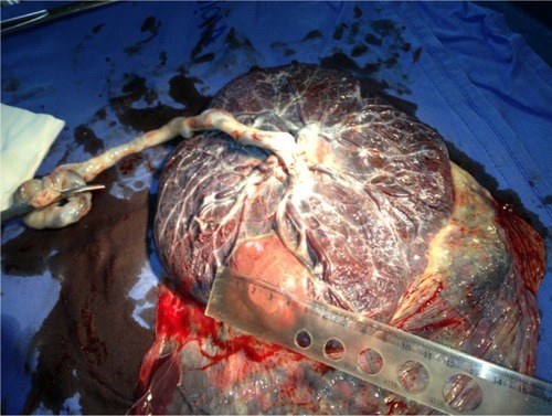
Figure 4 The atypical marginal location of the placental chorioangioma near the placental edge.
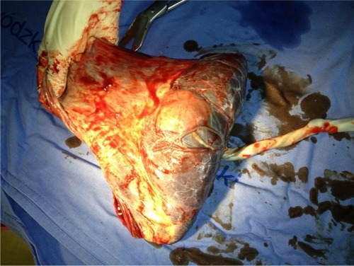
Figure 5 The complete maternal side of the placenta.